 Explore
 Validate
 Learn
Learn Western blot
Antibody data
- Antibody Data
- Antigen structure
- References [0]
- Comments [0]
- Validations
- Western blot [1]
- Immunohistochemistry [1]
Submit
Validation data
Reference
Comment
Report error
- Product number
- AF6539 - Provider product page

- Provider
- Novus Biologicals
- Product name
- Sheep Polyclonal ASCL2/Mash2 Antibody
- Antibody type
- Polyclonal
- Description
- Immunogen affinity purified. Detects human ASCL2/Mash2 in Western blots.
- Reactivity
- Human
- Host
- Sheep
- Isotype
- IgG
- Vial size
- 100 ug
- Concentration
- LYOPH
- Storage
- Use a manual defrost freezer and avoid repeated freeze-thaw cycles. 12 months from date of receipt, -20 to -70 degreesC as supplied. 1 month, 2 to 8 degreesC under sterile conditions after reconstitution. 6 months, -20 to -70 degreesC under sterile conditions after reconstitution.
No comments: Submit comment
Supportive validation
- Submitted by
- Novus Biologicals (provider)
- Main image
- Experimental details
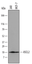
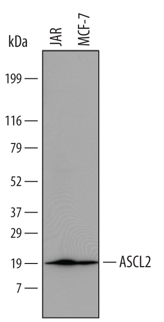
- Detection of Human ASCL2/Mash2 by Western Blot. Western blot shows lysates of JAR human choriocarcinoma cell line and MCF-7 human breast cancer cell line. PVDF Membrane was probed with 1 µg/mL of Sheep Anti-Human ASCL2/Mash2 Antigen Affinity-purified Polyclonal Antibody (Catalog # AF6539) followed by HRP-conjugated Anti-Sheep IgG Secondary Antibody (Catalog # HAF016). A specific band was detected for ASCL2/Mash2 at approximately 20 kDa (as indicated). This experiment was conducted under reducing conditions and using Immunoblot Buffer Group 1.
Supportive validation
- Submitted by
- Novus Biologicals (provider)
- Main image
- Experimental details
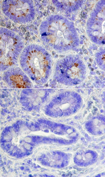
- ASCL2/Mash2 in Human Intestine. ASCL2/Mash2 was detected in immersion fixed paraffin-embedded sections of human intestine using Sheep Anti-Human ASCL2/Mash2 Antigen Affinity-purified Polyclonal Antibody (Catalog # AF6539) at 15 µg/mL overnight at 4 °C. Tissue was stained using the Anti-Sheep HRP-DAB Cell & Tissue Staining Kit (brown; Catalog # CTS019) and counterstained with hemotoxylin (blue). Lower panel shows a lack of labeling when primary antibodies are omitted and tissue is stained only with secondary antibody followed by incubation with detection reagents. Specific staining was localized to the base of intestinal crypts. View our protocol for Chromogenic IHC Staining of Paraffin-embedded Tissue Sections.
