Explore
 Validate
 Learn
Learn Western blot
Antibody data
- Antibody Data
- Antigen structure
- References [0]
- Comments [0]
- Validations
- Western blot [1]
- Immunocytochemistry [1]
Submit
Validation data
Reference
Comment
Report error
- Product number
- PA5-41489 - Provider product page

- Provider
- Invitrogen Antibodies
- Product name
- ASCL2 Polyclonal Antibody
- Antibody type
- Polyclonal
- Antigen
- Synthetic peptide
- Description
- Peptide sequence: RPQAVRPSAP RGPPGTTPVA ASPSRASSSP GRGGSSEPGS PRSAYSSDDS Sequence homology: Cow: 100%; Dog: 100%; Guinea Pig: 93%; Human: 100%; Mouse: 79%; Pig: 100%; Rat: 93%
- Reactivity
- Human
- Host
- Rabbit
- Isotype
- IgG
- Vial size
- 100 µL
- Concentration
- 0.5 mg/mL
- Storage
- -20° C, Avoid Freeze/Thaw Cycles
No comments: Submit comment
Supportive validation
- Submitted by
- Invitrogen Antibodies (provider)
- Main image
- Experimental details
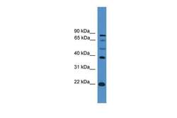
- Western blot analysis of human PANC1 cell lysate using an anti-ASCL2 polyclonal antibody (Product # PA5-41489).

Supportive validation
- Submitted by
- Invitrogen Antibodies (provider)
- Main image
- Experimental details
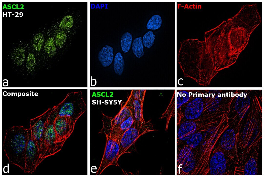
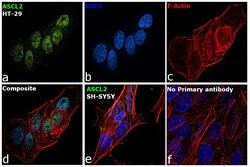
- Immunofluorescence analysis of ASCL2 was performed using HT-29 cells. The cells were fixed with 4% paraformaldehyde for 10 minutes, permeabilized with 0.1% Triton™ X-100 for 15 minutes, and blocked with 2% BSA for 1 hour at room temperature. The cells were labeled with ASCL2 Polyclonal Antibody (Product # PA5-41489) at 5 µg/mL in 0.1% BSA and incubated overnight at 4 degree and then labeled with Goat anti-Rabbit IgG (H+L) Superclonal™ Secondary Antibody, Alexa Fluor® 488 conjugate (Product # A27034) at a dilution of 1:2000 for 45 minutes at room temperature (Panel a: green). Nuclei (Panel b: blue) were stained with ProLong™ Diamond Antifade Mountant with DAPI (Product # P36962). F-actin (Panel c: red) was stained with Rhodamine Phalloidin (Product # R415, 1:300). Panel d represents the composite image showing nuclear localization of ASCL2. Panel e shows the negative model SH-SY5Y with no ASCL2 expression. Panel f represents control cells with no primary antibody to assess background. The images were captured at 60X magnification.