 Explore
 Validate
 Learn
Learn Western blot
Antibody data
- Antibody Data
- Antigen structure
- References [0]
- Comments [0]
- Validations
- Western blot [2]
- Immunohistochemistry [1]
Submit
Validation data
Reference
Comment
Report error
- Product number
- PA5-47852 - Provider product page

- Provider
- Invitrogen Antibodies
- Product name
- ASCL2 Polyclonal Antibody
- Antibody type
- Polyclonal
- Antigen
- Recombinant full-length protein
- Description
- Reconstitute in sterile PBS to a final concentration of 0.2 mg/mL.
- Reactivity
- Human
- Host
- Sheep
- Isotype
- IgG
- Vial size
- 100 µg
- Concentration
- 0.2 mg/mL
- Storage
- -20° C, Avoid Freeze/Thaw Cycles
No comments: Submit comment
Supportive validation
- Submitted by
- Invitrogen Antibodies (provider)
- Main image
- Experimental details
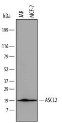
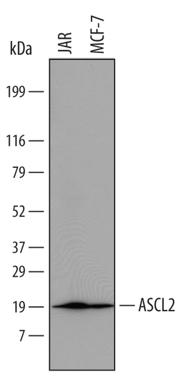
- Western blot analysis from lysates of JAR human choriocarcinoma cell line and MCF-7 human breast cancer cell line. PVDF Membrane was probed with 1 µg/mL of Sheep Anti-human ASCL2/Mash2 Antigen Affinity-purified Polyclonal Antibody (Product # PA5-47852) followed by HRP-conjugated Anti-Sheep IgG Secondary Antibody. A specific band was detected for ASCL2/Mash2 at approximately 20 kDa (as indicated). This experiment was conducted under reducing conditions.
- Submitted by
- Invitrogen Antibodies (provider)
- Main image

- Experimental details
- Western blot analysis of ASCL2 in JAR human choriocarcinoma cell line and MCF‚7 human breast cancer cell line. Samples were incubated in ASCL2 polyclonal antibody (Product # PA5-47852) using a dilution of 1 µg/mL followed by a HRP-conjugated Anti-Sheep IgG secondary antibody. A specific band was detected for ASCL2/Mash2 at approximately 20 kDa (as indicated). This experiment was conducted under reducing conditions.

Supportive validation
- Submitted by
- Invitrogen Antibodies (provider)
- Main image

- Experimental details
- Immunohistochemical analysis of ASCL2 in immersion fixed paraffin-embedded sections of human intestine. Samples were incubated in ASCL2 polyclonal antibody (Product # PA5-47852) using a dilution of 15 µg/mL overnight at 4 °C. Tissue was stained using the Anti-Sheep HRP-DAB Cell & Tissue Staining Kit (brown) and counterstained with hemotoxylin (blue). Lower panel shows a lack of labeling when primary antibodies are omitted and tissue is stained only with secondary antibody followed by incubation with detection reagents. Specific staining was localized to the base of intestinal crypts.
