 Explore
 Validate
 Learn
Learn Western blot
Antibody data
- Antibody Data
- Antigen structure
- References [0]
- Comments [0]
- Validations
- Western blot [2]
- Immunohistochemistry [2]
- Flow cytometry [2]
Submit
Validation data
Reference
Comment
Report error
- Product number
- MA1-46422 - Provider product page

- Provider
- Invitrogen Antibodies
- Product name
- LMO2 Monoclonal Antibody (1A9-3B11)
- Antibody type
- Monoclonal
- Antigen
- Recombinant full-length protein
- Description
- Suggested positive control: OCIL-Y19, Raji or Ramos whole cell lysates.
- Antibody clone number
- 1A9-3B11
- Concentration
- 1 mg/mL
No comments: Submit comment
Supportive validation
- Submitted by
- Invitrogen Antibodies (provider)
- Main image

- Experimental details
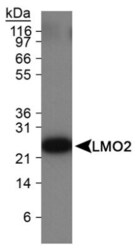
- Western Blot detection of LMO2 in Ramos cell lysate

- Submitted by
- Invitrogen Antibodies (provider)
- Main image
- Experimental details
- Western blot analysis of LMO2 in Ramos cell lysate. Sample was incubated in LMO2 monoclonal antibody (Product # MA1-46422).

Supportive validation
- Submitted by
- Invitrogen Antibodies (provider)
- Main image
- Experimental details
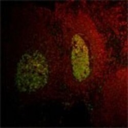
- Immunohistochemistry analysis of LMO2 in tonsil tissue. Samples were incubated in LMO2 monoclonal antibody (Product # MA1-46422). LMO2 (green).

- Submitted by
- Invitrogen Antibodies (provider)
- Main image

- Experimental details
- Immunohistochemical analysis of LMO2 in formalin fixed paraffin-embedded (FFPE) human tonsil. Samples were incubated in LMO2 monoclonal antibody (Product # MA1-46422) using a dilution of 1:500. Bond Rx autostainer (Leica Biosystems). The assay involved 20 minutes of heat induced antigen retrieval (HIER) using 10mM sodium citrate buffer (pH 6.0) and endogenous peroxidase quenching with peroxide block. The sections were incubated with primary antibody for 30 minutes and Bond Polymer Refine Detection (Leica Biosystems) with DAB was used for signal development followed by counterstaining with hematoxylin. Whole slide scanning and capturing of representative images was performed using Aperio AT2 (Leica Biosystems). Nuclear with some cytoplasmic staining was observed. Staining was performed by Histowiz.

Supportive validation
- Submitted by
- Invitrogen Antibodies (provider)
- Main image
- Experimental details
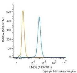
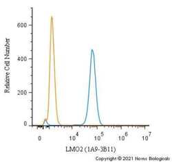
- Flow cytometry of LMO2 in THP-1 cells. Samples were incubated with LMO2 monoclonal antibody (Product # MA1-46422) using a dilution of 1.0 µg/mL for 30 minutes at room temperature followed by Mouse IgG (H+L) Cross-Adsorbed Secondary Antibody, Dylight 550 (Product # 35503). Antibody (blue) and a matched isotype control (orange). Cells were fixed with 4% PFA and then permeabilized with 0.1% saponin.
- Submitted by
- Invitrogen Antibodies (provider)
- Main image
- Experimental details
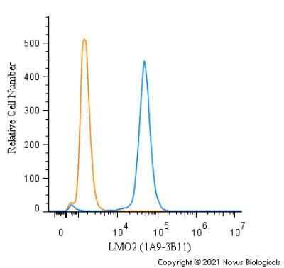
- Flow cytometry of LMO2 in Ramos cells. Samples were incubated with LMO2 monoclonal antibody (Product # MA1-46422) using a dilution of 1.0 µg/mL for 30 minutes at room temperature followed by Mouse IgG (H+L) Cross-Adsorbed Secondary Antibody, Dylight 550 (Product # 35503). Antibody (blue) and a matched isotype control (orange). Cells were fixed with 4% PFA and then permeabilized with 0.1% saponin.
