Explore
 Validate
 Learn
Learn Western blot
Antibody data
- Antibody Data
- Antigen structure
- References [1]
- Comments [0]
- Validations
- Western blot [1]
- Immunocytochemistry [2]
- Immunohistochemistry [1]
Submit
Validation data
Reference
Comment
Report error
- Product number
- 701585 - Provider product page

- Provider
- Invitrogen Antibodies
- Product name
- Troponin I Recombinant Rabbit Monoclonal Antibody (1H11L19)
- Antibody type
- Monoclonal
- Antigen
- Other
- Description
- Recombinant rabbit monoclonal antibodies are produced using in vitro expression systems. The expression systems are developed by cloning in the specific antibody DNA sequences from immunoreactive rabbits. Then, individual clones are screened to select the best candidates for production. The advantages of using recombinant rabbit monoclonal antibodies include: better specificity and sensitivity, lot-to-lot consistency, animal origin-free formulations, and broader immunoreactivity to diverse targets due to larger rabbit immune repertoire.
- Reactivity
- Human, Mouse, Rat
- Host
- Rabbit
- Isotype
- IgG
- Antibody clone number
- 1H11L19
- Vial size
- 100 µg
- Concentration
- 0.5 mg/mL
- Storage
- Store at 4°C short term. For long term storage, store at -20°C, avoiding freeze/thaw cycles.
Submitted references Metabolic Maturation Media Improve Physiological Function of Human iPSC-Derived Cardiomyocytes.
Feyen DAM, McKeithan WL, Bruyneel AAN, Spiering S, Hörmann L, Ulmer B, Zhang H, Briganti F, Schweizer M, Hegyi B, Liao Z, Pölönen RP, Ginsburg KS, Lam CK, Serrano R, Wahlquist C, Kreymerman A, Vu M, Amatya PL, Behrens CS, Ranjbarvaziri S, Maas RGC, Greenhaw M, Bernstein D, Wu JC, Bers DM, Eschenhagen T, Metallo CM, Mercola M
Cell reports 2020 Jul 21;32(3):107925
Cell reports 2020 Jul 21;32(3):107925
No comments: Submit comment
Supportive validation
- Submitted by
- Invitrogen Antibodies (provider)
- Main image
- Experimental details
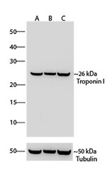
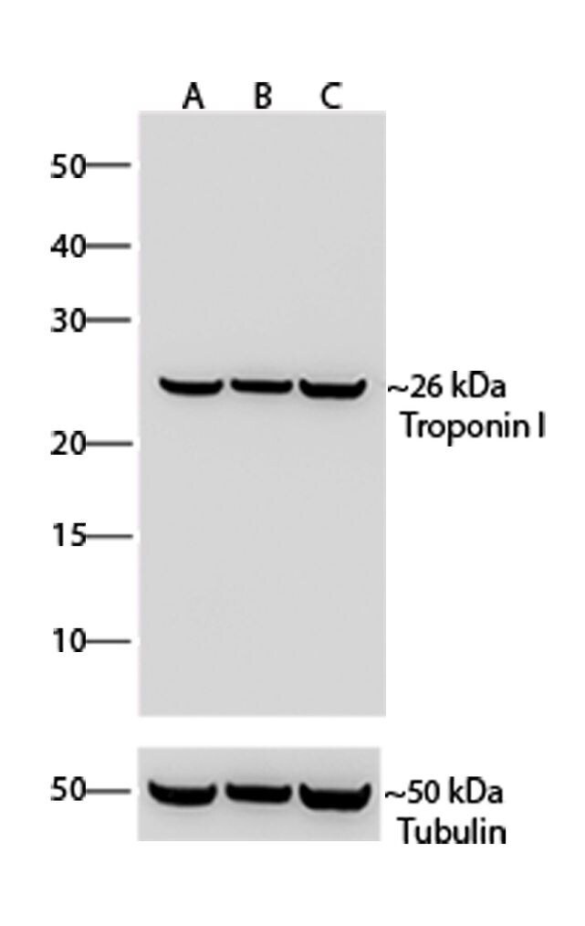
- Western blot analysis of Troponin I was performed by loading 20 µg of Mouse Heart, Rat Heart and Human Cardiomyocyte lysates (lane A, B, C) using Novex®NuPAGE®4-12% Bis-Tris gel (Product # NP0321BOX), Xcell SureLock Electrophoresis system (Product # EI0002), Novex sharp Pre-stained Protein Standard (Product # LC5800), and iBlot® Dry Blotting System (Product # IB21001). Proteins were transferred to a nitrocellulose membrane and blocked with 5% skim milk for 1 hour at room temperature. Troponin I was detected at ~26 kDa using Troponin I Recombinant Rabbit Monoclonal Antibody (Product # 701585) at a 1:1,000 dilution in 2.5% skim milk at 4° C overnight on a rocking platform. Goat anti-Rabbit IgG - HRP Secondary Antibody (Product # G-21234) at 1:5,000 dilution was used and chemiluminescent detection was performed using Pierce™ ECL Western blotting Substrate (Product # 32106).
Supportive validation
- Submitted by
- Invitrogen Antibodies (provider)
- Main image

- Experimental details
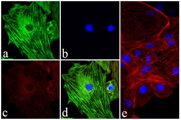
- Immunofluorescent analysis of Troponin I was done on Primary Human Cardiomyocytes. The cells were fixed with 4% paraformaldehyde for 15 minutes, permeabilized with 0.25% Triton™ X-100 for 10 minutes, and blocked with 5% BSA for 1 hour at room temperature. The cells were labeled with Troponin I Recombinant Rabbit Monoclonal Antibody (Product # 701585) at a dilution of 1:400 in 1% BSA and incubated for 3 hours at room temperature and then labeled with Alexa Fluor® 488 Goat anti-Rabbit IgG Secondary Antibody (Product # A-11008) at a dilution of 1:400 for 30 minutes at room temperature (Panel a: green). Nuclei (Panel b: blue) were stained with SlowFade® Gold Antifade Mountant with DAPI (Product # S36938). F-actin (Panel c: red) was stained with Alexa Fluor® 594 Phalloidin (Product # A12381). Panel d is a merged image showing striated muscle fiber localization. Panel e is a no primary antibody control. The images were captured using a Nikon microscope at 20X magnification.
- Submitted by
- Invitrogen Antibodies (provider)
- Main image
- Experimental details
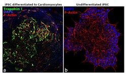
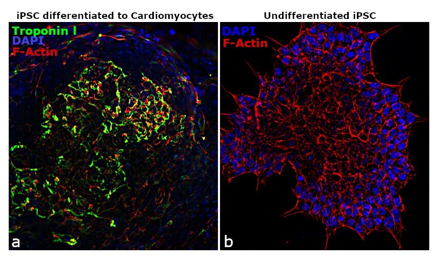
- For immunofluorescence analysis, iPSC differentiated to Cardiomyocytes were fixed and permeabilized for detection of endogenous Troponin I using anti-Troponin I Recombinant Rabbit monoclonal Antibody (Product # 701585, 1:100 dilution) and labeled with Goat anti-Rabbit IgG (H+L) Superclonal™ Secondary Antibody, Alexa Fluor® 488 conjugate (Product # A27034, 1:2,000).Nuclei (blue) is stained using ProLong™ Diamond Antifade Mountant with DAPI (Product # P36962). Panel c)and cytoskeletal F-actin stained using Rhodamine Phalloidin (Product # R415, 1:300). Panel a) shows undifferentiated iPSC cells, Panel b) is the image of differentiated cardiomyocytes showing cytoskeletal localization of sarcomeric Troponin I.
Supportive validation
- Submitted by
- Invitrogen Antibodies (provider)
- Main image

- Experimental details
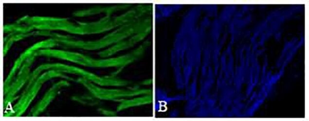
- Immunohistochemistry analysis of Troponin I Recombinant Rabbit Monoclonal Antibody (Product # 701585) was done on paraffin embedded mouse heart tissue sections. To expose target proteins, heat induced epitope retrieval was performed using Tris-EDTA (pH 9.0) buffer for 15 minutes. Following antigen retrieval, tissues were blocked in 0.2% BSA with 0.1% cold fish skin gelatin in 1X PBS for 1 hour in a humidified chamber. The tissues were then probed at a dilution of 1:100 with Troponin I Recombinant Rabbit Monoclonal Antibody (Product # 701585) or blocking buffer alone as negative control for 3 hours at room temperature in a humidified chamber. Detection was performed using Alexa Fluor® 488 Goat anti-Rabbit IgG Secondary Antibody (Product # A-11008) at a dilution of 1:400 for 30 minutes in a humidified chamber. Tissues were counterstained with SlowFade® Gold Antifade Mountant with DAPI (Product # S36938). The images were captured using a Nikon microscope at 20X magnification.