 Explore
 Validate
 Learn
LearnPA5-30292
antibody from Invitrogen Antibodies
Targeting: EIF2B4
DKFZP586J0119, EIF-2B, EIF2B, EIF2Bdelta
 Western blot
Antibody data
- Antibody Data
- Antigen structure
- References [0]
- Comments [0]
- Validations
- Western blot [3]
- Immunocytochemistry [2]
Submit
Validation data
Reference
Comment
Report error
- Product number
- PA5-30292 - Provider product page

- Provider
- Invitrogen Antibodies
- Product name
- eIF2b delta Polyclonal Antibody
- Antibody type
- Polyclonal
- Antigen
- Recombinant protein fragment
- Description
- Recommended positive controls: 293T, A431, Raji.
- Concentration
- 0.61 mg/mL
No comments: Submit comment
Supportive validation
- Submitted by
- Invitrogen Antibodies (provider)
- Main image
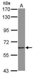
- Experimental details
- Western Blot using eIF2b delta Polyclonal Antibody (Product # PA5-30292). Sample (30 µg of whole cell lysate). Lane A: A431 . 7.5% SDS PAGE. eIF2b delta Polyclonal Antibody (Product # PA5-30292) diluted at 1:1,000.

- Submitted by
- Invitrogen Antibodies (provider)
- Main image

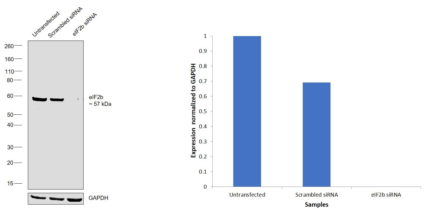
- Experimental details
- Knockdown of Translation initiation factor eIF-2B subunit delta was achieved by transfecting HEK-293 with Translation initiation factor eIF-2B subunit delta specific siRNAs (Silencer® select Product # s531804, s16992). Western Blot analysis (Fig. a) was performed using Whole cell extracts from the Translation initiation factor eIF-2B subunit delta knockdown cells (lane 3), non-targeting scrambled siRNA transfected cells (lane 2) and untransfected cells (lane 1). The Blot was probed with eIF2b delta Polyclonal Antibody (Product # PA5-30292, 1:1000 dilution ) and Goat anti-Rabbit IgG (H+L) Superclonal™ Recombinant Secondary Antibody, HRP (Product # A27036, 1:10,000 dilution). Densitometric analysis of this western Blot is shown in histogram (Fig. b). Decrease in signal upon siRNA mediated knock down confirms that antibody is specific to Translation initiation factor eIF-2B subunit delta.
- Submitted by
- Invitrogen Antibodies (provider)
- Main image
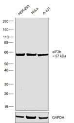
- Experimental details
- Western Blot was performed using Anti-eIF2b delta Polyclonal Antibody (Product # PA5-30292) and a 57 kDa band corresponding to Translation initiation factor eIF-2B subunit delta was observed across across the cell lines tested. Whole cell extracts (30 µg lysate) of HEK-293 (Lane 1), HeLa (Lane 2), A-431 (Lane 3) were electrophoresed using NuPAGE™ 4-12% Bis-Tris Protein Gel (Product # NP0321BOX). Resolved proteins were then transferred onto a Nitrocellulose membrane (Product # IB23001) by iBlot® 2 Dry Blotting System (Product # IB21001). The Blot was probed with the primary antibody (1:1000 diluion) and detected by chemiluminescence with Goat anti-Rabbit IgG (H+L) Superclonal™ Recombinant Secondary Antibody, HRP (Product # A27036, 1:10000 dilution) using the iBright FL 1000 (Product # A32752). Chemiluminescent detection was performed using Novex® ECL Chemiluminescent Substrate Reagent Kit (Product # WP20005).

Supportive validation
- Submitted by
- Invitrogen Antibodies (provider)
- Main image
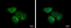
- Experimental details
- eIF2b delta Polyclonal Antibody detects EIF2B delta protein at cytoplasm by immunofluorescent analysis. Sample: A431 cells were fixed in 4% paraformaldehyde at RT for 15 min. Green: EIF2B delta protein stained by eIF2b delta Polyclonal Antibody (Product # PA5-30292) diluted at 1:500. Blue: Hoechst 33342 staining.

- Submitted by
- Invitrogen Antibodies (provider)
- Main image
- Experimental details
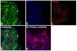
- Immunofluorescence analysis of Translation initiation factor eIF-2B subunit delta was performed using 70% confluent log phase A-431 cells. The cells were fixed with 4% paraformaldehyde for 10 minutes, permeabilized with 0.1% Triton™ X-100 for 15 minutes, and blocked with 2% BSA for 45 minutes at room temperature. The cells were labeled with eIF2b delta Polyclonal Antibody (Product # PA5-30292) at 1:100 dilution in 0.1% BSA, incubated at 4 degree celsius overnight and then labeled with Goat anti-Rabbit IgG (H+L) Superclonal™ Recombinant Secondary Antibody, Alexa Fluor® 488 conjµgate (Product # A27034), (1:2000 dilution), for 45 minutes at room temperature (Panel a: Green). Nuclei (Panel b:Blue) were stained with ProLong™ Diamond Antifade Mountant with DAPI (Product # P36962). F-actin (Panel c: Red) was stained with Rhodamine Phalloidin (Product # R415, 1:300). Panel d represents the merged image showing cytoplasmic localization. Panel e represents control cells with no primary antibody to assess background. The images were captured at 40X magnification.
