 Explore
 Validate
 Learn
Learn Western blot
Antibody data
- Antibody Data
- Antigen structure
- References [0]
- Comments [0]
- Validations
- Western blot [3]
- Immunocytochemistry [2]
- Immunohistochemistry [7]
Submit
Validation data
Reference
Comment
Report error
- Product number
- PA5-53922 - Provider product page

- Provider
- Invitrogen Antibodies
- Product name
- Nuclear Matrix Protein p84 Polyclonal Antibody
- Antibody type
- Polyclonal
- Antigen
- Recombinant full-length protein
- Description
- Immunogen sequence: VFCGRIQLFL ARLFPLSEKS GLNLQSQFNL ENVTVFNTNE QESTLGQKHT EDREEGMDVE EGEMGDEEAP TTCSIPIDYN LYRKFWS Highest antigen sequence identity to the following orthologs: Mouse - 99%, Rat - 46%.
- Reactivity
- Human
- Host
- Rabbit
- Isotype
- IgG
- Vial size
- 100 µL
- Concentration
- 0.1 mg/mL
- Storage
- Store at 4°C short term. For long term storage, store at -20°C, avoiding freeze/thaw cycles.
No comments: Submit comment
Supportive validation
- Submitted by
- Invitrogen Antibodies (provider)
- Main image

- Experimental details
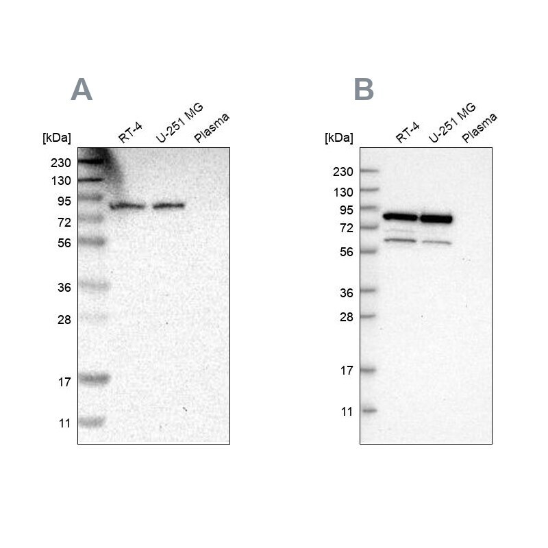
- Western blot analysis of Nuclear Matrix Protein p84 using Nuclear Matrix Protein p84 Polyclonal Antibody (Product # PA5-53922) (A) shows similar pattern to an independent Nuclear Matrix Protein p84 Polyclonal Antibody (B).
- Submitted by
- Invitrogen Antibodies (provider)
- Main image

- Experimental details
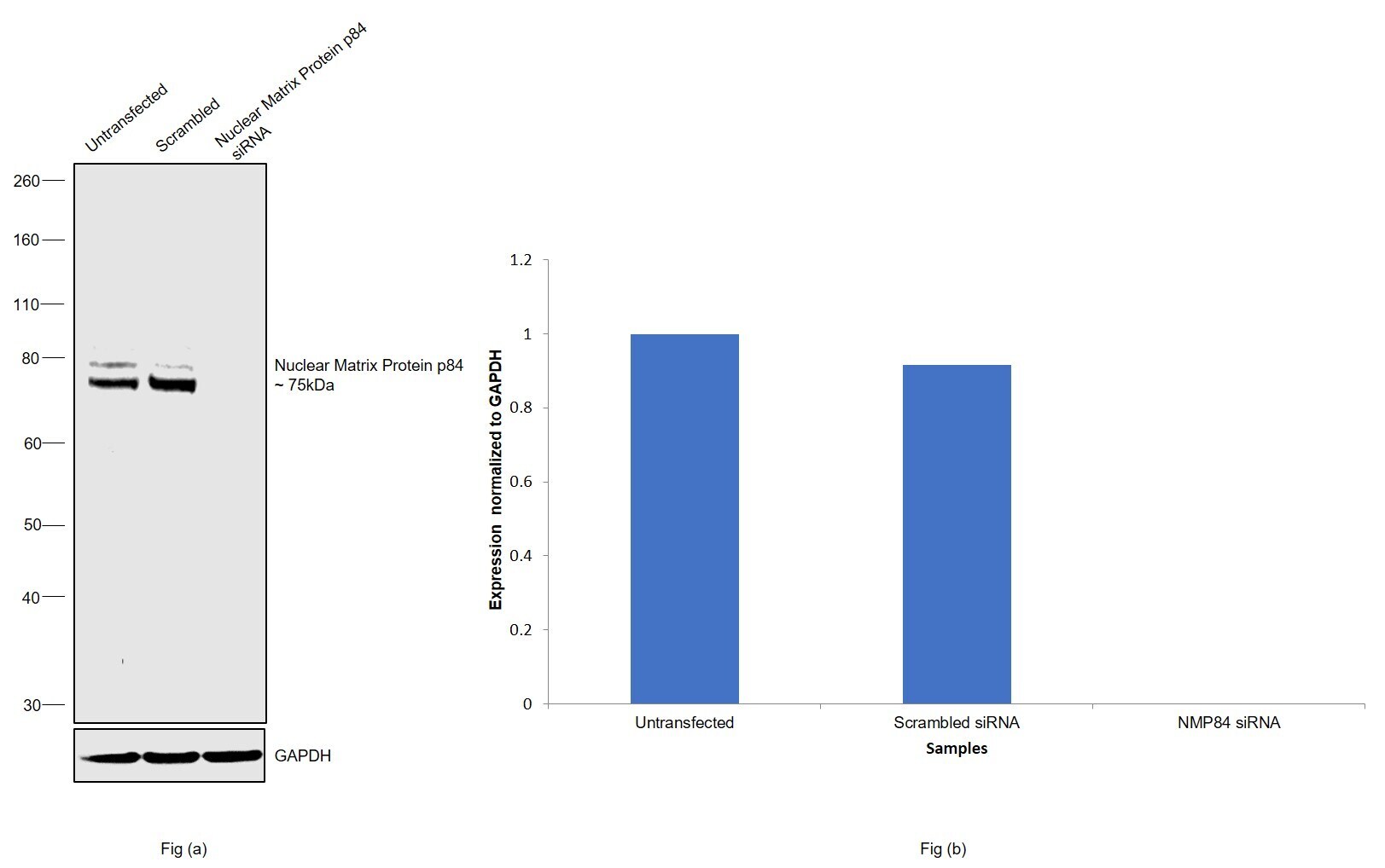
- Knockdown of Nuclear Matrix Protein p84 was achieved by transfecting HeLa with Nuclear Matrix Protein p84 specific siRNAs (Silencer® select Product # s19396, s19397). Western blot analysis (Fig. a) was performed using whole cell extracts from the Nuclear Matrix Protein p84 knockdown cells (lane 3), non-specific scrambled siRNA transfected cells (lane 2) and untransfected cells (lane 1). The blot was probed with Nuclear Matrix Protein p84 Polyclonal Antibody (Product # PA5-53922, 0.2ug/ml) and Goat anti-Rabbit IgG (H+L) Superclonal™ Secondary Antibody, HRP (Product # A27036, 0.25µg/ml, 1:4000 dilution). Densitometric analysis of this western blot is shown in histogram (Fig. b). Decrease in signal upon siRNA mediated knock down confirms that antibody is specific to Nuclear Matrix Protein p84.
- Submitted by
- Invitrogen Antibodies (provider)
- Main image

- Experimental details
- Western blot was performed using anti-Nuclear Matrix Protein p84 Polyclonal Antibody (Product # PA5-53922) and a 75 kDa band corresponding to Nuclear Matrix Protein p84 was observed across all the cell lines and tissues tested. Whole cell extracts (30 µg lysate) of HeLa (Lane 1), A549 (Lane 2), MDA-MB-231 (Lane 3), MCF 10A (Lane 4), tissue extracts of Mouse Testis (Lane 5) and Mouse Skeletal Muscle (Lane 6) were electrophoresed using NuPAGE™ 4-12% Bis-Tris Protein Gel (Product # NP0322BOX). Relative expression of Nuclear Matrix Protein p84 Polyclonal Antibody was observed high in Mouse testis and low or negative in Mouse skeletal Muscle. Resolved proteins were then transferred onto a nitrocellulose membrane (Product # IB23001) by iBlot® 2 Dry Blotting System (Product # IB21001). The blot was probed with the primary antibody (0.2 µg/mL) and detected by chemiluminescence with Goat anti-Rabbit IgG (H+L), Superclonal™ Recombinant Secondary Antibody, HRP (Product # A27036, 1:4000 dilution) using the iBright FL 1000 (Product # A32752). Chemiluminescent detection was performed using Novex® ECL Chemiluminescent Substrate Reagent Kit (Product # WP20005).

Supportive validation
- Submitted by
- Invitrogen Antibodies (provider)
- Main image
- Experimental details
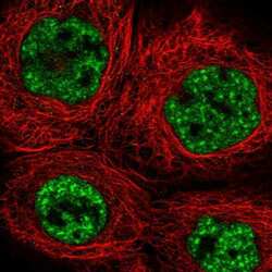
- Immunofluorescent staining of Nuclear Matrix Protein p84 in human cell line A-431 shows positivity in nucleus but excluded from the nucleoli. Samples were probed using a Nuclear Matrix Protein p84 Polyclonal Antibody (Product # PA5-53922).

- Submitted by
- Invitrogen Antibodies (provider)
- Main image

- Experimental details
- Immunofluorescence analysis of Nuclear Matrix Protein p84 was performed using 70% confluent log phase HeLa cells. The cells were fixed with 4% paraformaldehyde for 10 minutes, permeabilized with 0.1% Triton™ X-100 for 15 minutes, and blocked with 2% BSA for 45 minutes at room temperature. The cells were labeled with Nuclear Matrix Protein p84 Polyclonal Antibody (Product # PA5-53922) at 5 µg/mL in 0.1% BSA, incubated at 4 degree celsius overnight and then labeled with Donkey anti-Rabbit IgG (H+L) Highly Cross-Adsorbed Secondary Antibody, Alexa Fluor Plus 488 (Product # A32790), (1:2000 dilution) for 45 minutes at room temperature (Panel a: Green). Nuclei (Panel b: Blue) were stained with SlowFade® Gold Antifade Mountant with DAPI (Product # S36938). F-actin (Panel c: Red) was stained with Rhodamine Phalloidin (Product # R415, 1:300 dilution). Panel d represents the merged image showing nuclear localization. Panel e represents control cells with no primary antibody to assess background. The images were captured at 60X magnification.

Supportive validation
- Submitted by
- Invitrogen Antibodies (provider)
- Main image

- Experimental details
- Immunohistochemical staining of Nuclear Matrix Protein p84 in human testis using Nuclear Matrix Protein p84 Polyclonal Antibody (Product # PA5-53922).

- Submitted by
- Invitrogen Antibodies (provider)
- Main image

- Experimental details
- Immunohistochemical staining of Nuclear Matrix Protein p84 in human colon using Nuclear Matrix Protein p84 Polyclonal Antibody (Product # PA5-53922).

- Submitted by
- Invitrogen Antibodies (provider)
- Main image

- Experimental details
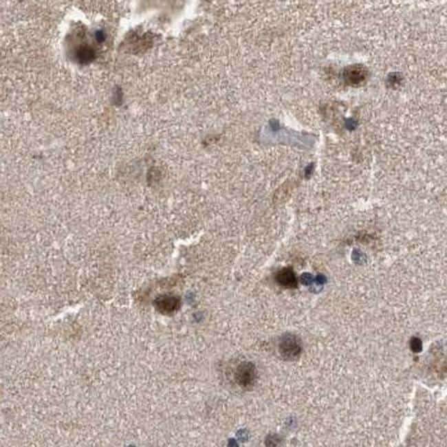
- Immunohistochemical staining of Nuclear Matrix Protein p84 in human cerebral cortex using Nuclear Matrix Protein p84 Polyclonal Antibody (Product # PA5-53922).
- Submitted by
- Invitrogen Antibodies (provider)
- Main image

- Experimental details
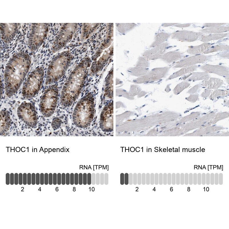
- Immunohistochemical staining of Nuclear Matrix Protein p84 in human appendix and skeletal muscle tissues using Nuclear Matrix Protein p84 Polyclonal Antibody (Product # PA5-53922). Corresponding THOC1 RNA-seq data are presented for the same tissues.
- Submitted by
- Invitrogen Antibodies (provider)
- Main image

- Experimental details
- Immunohistochemical staining of Nuclear Matrix Protein p84 in human cerebral cortex, colon, skeletal muscle and testis using Nuclear Matrix Protein p84 Polyclonal Antibody (Product # PA5-53922) (A) shows similar protein distribution across tissues to an independent Nuclear Matrix Protein p84 Polyclonal Antibody (B).

- Submitted by
- Invitrogen Antibodies (provider)
- Main image

- Experimental details
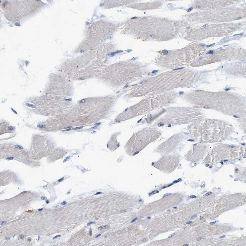
- Immunohistochemical staining of Nuclear Matrix Protein p84 in human skeletal muscle using Nuclear Matrix Protein p84 Polyclonal Antibody (Product # PA5-53922) shows low expression as expected.
- Submitted by
- Invitrogen Antibodies (provider)
- Main image
- Experimental details
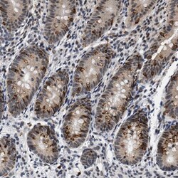
- Immunohistochemical staining of Nuclear Matrix Protein p84 in human appendix using Nuclear Matrix Protein p84 Polyclonal Antibody (Product # PA5-53922) shows high expression.
