Explore
 Validate
 Learn
Learn Western blot
Antibody data
- Antibody Data
- Antigen structure
- References [0]
- Comments [0]
- Validations
- Western blot [1]
- Immunohistochemistry [1]
Submit
Validation data
Reference
Comment
Report error
- Product number
- MAB3838 - Provider product page

- Provider
- R&D Systems
- Product name
- Rat Serpin A6 Antibody
- Antibody type
- Monoclonal
- Description
- Protein A or G purified from hybridoma culture supernatant. Detects rat Serpin A6 in direct ELISAs.
- Reactivity
- Rat
- Host
- Mouse
- Conjugate
- Unconjugated
- Antigen sequence
P31211- Isotype
- IgG
- Antibody clone number
- 930816
- Vial size
- 100 ug
- Storage
- Use a manual defrost freezer and avoid repeated freeze-thaw cycles. 12 months from date of receipt, -20 to -70 °C as supplied. 1 month, 2 to 8 °C under sterile conditions after reconstitution. 6 months, -20 to -70 °C under sterile conditions after reconstitution.
No comments: Submit comment
Supportive validation
- Submitted by
- R&D Systems (provider)
- Main image
- Experimental details
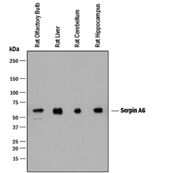
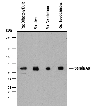
- Detection of Rat Serpin A6 by Western Blot. Western blot shows lysates of rat olfactory bulb tissue, rat liver tissue, rat brain (cerebellum) tissue, and rat brain (hippocampus) tissue. PVDF membrane was probed with 0.2 µg/mL of Mouse Anti-Rat Serpin A6 Monoclonal Antibody (Catalog # MAB3838) followed by HRP-conjugated Anti-Mouse IgG Secondary Antibody (Catalog # HAF018). A specific band was detected for Serpin A6 at approximately 50-60 kDa (as indicated). This experiment was conducted under reducing conditions and using Immunoblot Buffer Group 1.
Supportive validation
- Submitted by
- R&D Systems (provider)
- Main image

- Experimental details
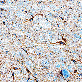
- Serpin A6 in Rat Brain. Serpin A6 was detected in perfusion fixed frozen sections of rat brain (preoptic nucleus) using Mouse Anti-Rat Serpin A6 Monoclonal Antibody (Catalog # MAB3838) at 25 µg/mL overnight at 4 °C. Tissue was stained using the Anti-Mouse HRP-DAB Cell & Tissue Staining Kit (brown; Catalog # CTS002) and counterstained with hematoxylin (blue). Specific staining was localized to neurons. View our protocol for Chromogenic IHC Staining of Frozen Tissue Sections.