Explore
 Validate
 Learn
LearnNBP2-52405
antibody from Novus Biologicals
Targeting: CTCFL
BORIS, CT27, dJ579F20.2
 Western blot
 ELISA Immunocytochemistry Immunoprecipitation Immunohistochemistry Chromatin Immunoprecipitation
Antibody data
- Antibody Data
- Antigen structure
- References [1]
- Comments [0]
- Validations
- Western blot [1]
- Immunoprecipitation [1]
- Immunohistochemistry [1]
Submit
Validation data
Reference
Comment
Report error
- Product number
- NBP2-52405 - Provider product page

- Provider
- Novus Biologicals
- Product name
- Mouse Monoclonal BORIS Antibody
- Antibody type
- Monoclonal
- Description
- Protein G purified.
- Reactivity
- Human
- Host
- Mouse
- Isotype
- IgG
- Vial size
- 0.1 ml
- Concentration
- 1 mg/ml
- Storage
- Store at 4C short term. Aliquot and store at -20C long term. Avoid freeze-thaw cycles.
Submitted references BORIS promotes chromatin regulatory interactions in treatment-resistant cancer cells.
Debruyne DN, Dries R, Sengupta S, Seruggia D, Gao Y, Sharma B, Huang H, Moreau L, McLane M, Day DS, Marco E, Chen T, Gray NS, Wong KK, Orkin SH, Yuan GC, Young RA, George RE
Nature 2019 Aug;572(7771):676-680
Nature 2019 Aug;572(7771):676-680
No comments: Submit comment
Supportive validation
- Submitted by
- Novus Biologicals (provider)
- Main image
- Experimental details
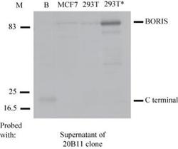
- Western Blot: BORIS Antibody (20B11) [NBP2-52405] - Antibody from clone 20B11 specifically recognises the C terminal domain of BORIS, and the endogenous and exogenous BORIS protein. Cell lysates were resolved by SDS-PAGE, blotted and probed with the original mouse supernatant of the 20B11 clone. Positions of BORIS and the C-terminal domain of BORIS are indicated. B, bacterially expressed BORIS; 293T cells transfected with 0.5ug of the plasmid expressing BORIS; M, Molecular marker.
Supportive validation
- Submitted by
- Novus Biologicals (provider)
- Main image

- Experimental details
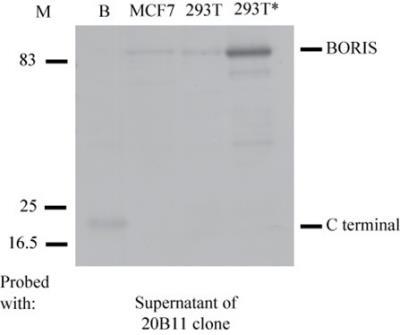
- Immunoprecipitation: BORIS Antibody (20B11) [NBP2-52405] - The 83-kDa protein can be immunoprecipitated with the mouse anti- BORIS C terminal specific antibody. Proteins were immunoprecipitated from the MCF7 cell extracts with the mouse anti- BORIS C terminal antibody 20B11 in their original supernatant form using the following volumes: (1) 400 ul of 20B11; (2) 800 ul of 20B11; (3) Input; (4) MCF7 cell lysates as BORIS positive control. The extracts from MCF7 cells were incubated overnight at 40C followed by 2 hour incubation with protein G sepharose beads. Proteins were resolved by SDS-PAGE, and Western blot analysis was performed with the concentrated form of the mouse anti- BORIS N terminal (4A7) specific antibody (1:500).

Supportive validation
- Submitted by
- Novus Biologicals (provider)
- Main image

- Experimental details
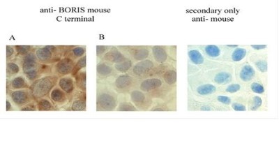
- Immunohistochemistry: BORIS Antibody (20B11) [NBP2-52405] - Analysis using the supernatants from the clone 20B11 at following dilutions: (A) 20B11 undiluted supernatant and (B) 20B11 at 1:25. (B) Cells have been counterstained with the nuclear marker hematoxylin (blue). Images were taken at x60 magnification using the Olympus BX41 microscope. Expression of BORIS protein is indicated by the presence of brown staining, with a pattern characteristic for BORIS ( both, cytoplasmic and nuclear).