Explore
 Validate
 Learn
Learn Western blot
Antibody data
- Antibody Data
- Antigen structure
- References [0]
- Comments [0]
- Validations
- Western blot [2]
- ELISA [1]
- Immunohistochemistry [1]
- Other assay [1]
Submit
Validation data
Reference
Comment
Report error
- Product number
- MA5-14680 - Provider product page

- Provider
- Invitrogen Antibodies
- Product name
- hCG Monoclonal Antibody (SC1)
- Antibody type
- Monoclonal
- Antigen
- Other
- Description
- The observed molecular weight of alpha-hCG and beta-hCG are approximately 20-22 kD & 27-32 kD respectively.
- Antibody clone number
- SC1
- Concentration
- 1 mg/mL
No comments: Submit comment
Supportive validation
- Submitted by
- Invitrogen Antibodies (provider)
- Main image
- Experimental details
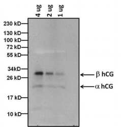
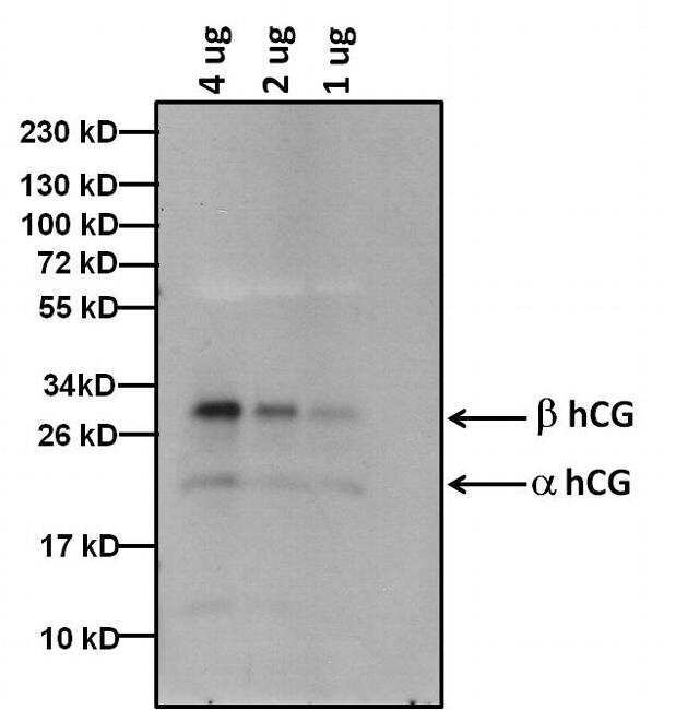
- Western blot analysis of hCG was performed by loading the indicated amounts of purified hCG protein extracted from human urine, and 15 µL of PageRuler Prestained Protein Ladder (Product # 26619) per well onto a 4-20% Tris-HCl polyacrylamide gel. Proteins were transferred to a PVDF membrane (Product # 88518) using the G2 Fast Blotter (Product # 62288) and blocked with 5% Milk for at least 1 hour at room temperature. hCG was detected at ~32 & 22 kD using a anti hCG antibody (Product # MA5-14680) at a concentration of 4 µg/mL in blocking buffer overnight at 4C on a rocking platform, followed by an HRP-conjugated goat anti-mouse IgG (Fc) secondary antibody (Product # 31439) at a dilution of 1:10,000 for at least 1 hour. Chemiluminescent detection was performed using SuperSignal West Dura (Product # 34076).
- Submitted by
- Invitrogen Antibodies (provider)
- Main image

- Experimental details
- Western blot analysis of hCG was performed by loading the indicated amounts of purified hCG protein extracted from human urine, and 15 µL of PageRuler Prestained Protein Ladder (Product # 26619) per well onto a 4-20% Tris-HCl polyacrylamide gel. Proteins were transferred to a PVDF membrane (Product # 88518) using the G2 Fast Blotter (Product # 62288) and blocked with 5% Milk for at least 1 hour at room temperature. hCG was detected at ~32 & 22 kD using a anti hCG antibody (Product # MA5-14680) at a concentration of 4 µg/mL in blocking buffer overnight at 4C on a rocking platform, followed by an HRP-conjugated goat anti-mouse IgG (Fc) secondary antibody (Product # 31439) at a dilution of 1:10,000 for at least 1 hour. Chemiluminescent detection was performed using SuperSignal West Dura (Product # 34076).

Supportive validation
- Submitted by
- Invitrogen Antibodies (provider)
- Main image
- Experimental details
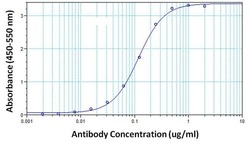
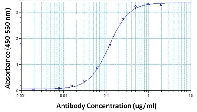
- Direct ELISA analysis of hCG proteins was performed by coating wells of a plate with a hCG protein extracted from human urine at a concentration of 10 µg/mL overnight at 4C. The plate was washed 3 times with ELISA Wash Buffer (Product # N503) and blocked with 300 µL of StartingBlock (PBS) Blocking Buffer (Product # 37538) for at least 1 hour at room temperature. After washing 100 µL of hCG monoclonal antibody (Product # MA5-14680) was added to wells in duplicate at 10, 5, 2.5, 1.25, 0.62, 0.31, 0.15, 9.5, 0.078, 0.039, 0.019 and 0 µg/mL concentrations, and the samples were incubated for 2 hours at room temperature. The plate was washed, and then incubated with 100 µL per well of an HRP-conjugated goat anti-mouse IgG secondary antibody (Product # 31430) at a dilution of 1:10,000 for 1 hour at room temperature, and washed again with ELISA Wash Buffer. The plate was developed by incubating 100 µL per well of 1-Step Ultra TMB substrate (Product # 34028) per well for 10 minutes at room temperature in the dark. The reaction was stopped with 100 µL per well of 0.16M sulfuric acid. Absorbances were read on a spectrophotometer at 450-550 nm.
Supportive validation
- Submitted by
- Invitrogen Antibodies (provider)
- Main image
- Experimental details
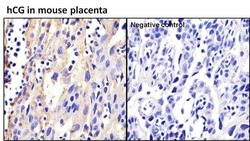
- Immunohistochemistry analysis of hCG was performed on human placenta tissue and is showing positive staining in extra cellular spaces. To expose target proteins, antigen retrieval was performed by microwaving tissues for 20 minutes in 10mM sodium citrate buffer (pH 6.0). Tissue slides were probed with hCG monoclonal antibody (Product # MA5-14680) at a dilution of 1:20, overnight at 4C in a humidified chamber without (right panel) or with hCG antibody (left panel). Tissues were washed, and incubated with secondary antibody (conjugated with HRP) for 30 min at room temperature in a humidified chamber. Detection was performed using a DAB substrate kit. Tissues were counterstained with hematoxylin and dehydrated to prep for mounting. Images were taken at 20x magnification.

Supportive validation
- Submitted by
- Invitrogen Antibodies (provider)
- Main image

- Experimental details
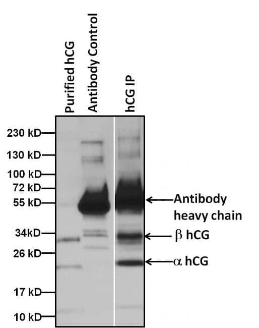
- Immunoprecipitation of hCG proteins was performed by spiking 5 ugs of purified hCG extracted from human urine into 1 mL of 1:2 diluted human urine sample. Antigen-antibody complexes were formed by incubating with 5 µg of hCG antibody (Product # MA5-14680) overnight on end over end tube rotator at 4C. The immune complexes were captured by using 50 µL Protein A/G Agarose (Product # 20421), washed extensively, and eluted with Lane Marker Reducing Sample Buffer (Product # 39000). Samples were resolved on a 4-20% Tris-HCl polyacrylamide gel, transferred to a PVDF membrane (Product # 88518), and blocked with 5% milk for at least 1 hour at room temperature. hCG was detected at ~32 & 22 kD using hCG monoclonal antibody (Product # MA5-14680) at a concentration of 4 µg/mL in blocking buffer overnight at 4C on a rocking platform, followed by an HRP-conjugated goat anti-mouse IgG (Fc) secondary antibody (Product # 31439) at a dilution of 1:10,000 for at least 1 hour. Chemiluminescent detection was performed using SuperSignal West Dura (Product # 34076).