Explore
 Validate
 Learn
Learn Western blot
 Immunohistochemistry
Antibody data
- Antibody Data
- Antigen structure
- References [0]
- Comments [0]
- Validations
- Immunohistochemistry [1]
Submit
Validation data
Reference
Comment
Report error
- Product number
- PB9234 - Provider product page

- Provider
- Boster Biological Technology
- Product name
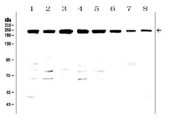
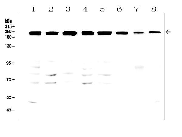
- Anti-ZO1 tight junction protein/TJP1 Antibody Picoband™
- Antibody type
- Polyclonal
- Description
- Polyclonal antibody for TIGHT JUNCTION PROTEIN 1/TJP1 detection. Host: Rabbit.Size: 100μg/vial. Tested applications: WB, IHC-P, ICC/IF, FCM. Reactive species: Human;Mouse;Rat. TIGHT JUNCTION PROTEIN 1/TJP1 information: Molecular Weight: 195459 MW; Subcellular Localization: Cell membrane; Peripheral membrane protein; Cytoplasmic side. Cell junction, tight junction. Cell junction. Cell junction, gap junction. Moves from the cytoplasm to the cell membrane concurrently with cell-cell contact. Detected at the leading edge of migrating and wounded cells; Tissue Specificity: The alpha-containing isoform is found in most epithelial cell junctions. The short isoform is found both in endothelial cells and the highly specialized epithelial junctions of renal glomeruli and Sertoli cells of the seminiferous tubules.
- Reactivity
- Human, Mouse, Rat
- Host
- Rabbit
- Vial size
- 100μg/vial
- Concentration
- Add 0.2ml of distilled water will yield a concentration of 500ug/ml.
- Storage
- At -20°C for one year. After reconstitution, at 4°C for one month. It can also be aliquoted and stored frozen at -20°C for a longer time. Avoid repeated freezing and thawing.
- Handling
- Add 0.2ml of distilled water will yield a concentration of 500ug/ml.
No comments: Submit comment
Supportive validation
- Submitted by
- Boster Biological Technology (provider)
- Main image
- Experimental details
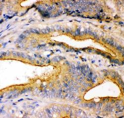
- IHC analysis of TJP1 using anti-TJP1 antibody (PB9234).TJP1 was detected in paraffin-embedded section of human intestinal cancer tissue. Heat mediated antigen retrieval was performed in citrate buffer (pH6, epitope retrieval solution) for 20 mins. The tissue section was blocked with 10% goat serum. The tissue section was then incubated with 1μg/ml rabbit anti-TJP1 Antibody (PB9234) overnight at 4°C. Biotinylated goat anti-rabbit IgG was used as secondary antibody and incubated for 30 minutes at 37°C. The tissue section was developed using Strepavidin-Biotin-Complex (SABC)(Catalog # SA1022) with DAB as the chromogen.
- Additional image