Explore
 Validate
 Learn
Learn Western blot
 Immunocytochemistry
 Immunoprecipitation
Antibody data
- Antibody Data
- Antigen structure
- References [5]
- Comments [0]
- Validations
- Western blot [6]
- Immunocytochemistry [3]
- Immunohistochemistry [3]
Submit
Validation data
Reference
Comment
Report error
- Product number
- GTX108613 - Provider product page

- Provider
- GeneTex
- Proper citation
- GeneTex Cat#GTX108613, RRID:AB_1952257
- Product name
- ZO-1 antibody [N1N2], N-term
- Antibody type
- Polyclonal
- Reactivity
- Human, Mouse
- Host
- Rabbit
Submitted references Astragaloside IV protects blood-brain barrier integrity from LPS-induced disruption via activating Nrf2 antioxidant signaling pathway in mice.
SLC26A3 (DRA) prevents TNF-alpha-induced barrier dysfunction and dextran sulfate sodium-induced acute colitis.
Neutralization of IL-8 decreases tumor PMN-MDSCs and reduces mesenchymalization of claudin-low triple-negative breast cancer.
Helicobacter pylori CagA and IL-1β Promote the Epithelial-to-Mesenchymal Transition in a Nontransformed Epithelial Cell Model.
The Synthetic β-Nitrostyrene Derivative CYT-Rx20 Inhibits Esophageal Tumor Growth and Metastasis via PI3K/AKT and STAT3 Pathways.
Li H, Wang P, Huang F, Jin J, Wu H, Zhang B, Wang Z, Shi H, Wu X
Toxicology and applied pharmacology 2018 Feb 1;340:58-66
Toxicology and applied pharmacology 2018 Feb 1;340:58-66
SLC26A3 (DRA) prevents TNF-alpha-induced barrier dysfunction and dextran sulfate sodium-induced acute colitis.
Ding X, Li D, Li M, Wang H, He Q, Wang Y, Yu H, Tian D, Yu Q
Laboratory investigation; a journal of technical methods and pathology 2018 Apr;98(4):462-476
Laboratory investigation; a journal of technical methods and pathology 2018 Apr;98(4):462-476
Neutralization of IL-8 decreases tumor PMN-MDSCs and reduces mesenchymalization of claudin-low triple-negative breast cancer.
Dominguez C, McCampbell KK, David JM, Palena C
JCI insight 2017 Nov 2;2(21)
JCI insight 2017 Nov 2;2(21)
Helicobacter pylori CagA and IL-1β Promote the Epithelial-to-Mesenchymal Transition in a Nontransformed Epithelial Cell Model.
Arévalo-Romero H, Meza I, Vallejo-Flores G, Fuentes-Pananá EM
Gastroenterology research and practice 2016;2016:4969163
Gastroenterology research and practice 2016;2016:4969163
The Synthetic β-Nitrostyrene Derivative CYT-Rx20 Inhibits Esophageal Tumor Growth and Metastasis via PI3K/AKT and STAT3 Pathways.
Chiu WC, Lee YC, Su YH, Wang YY, Tsai CH, Hou YA, Wang CH, Huang YF, Huang CJ, Chou SH, Hsieh PW, Yuan SF
PloS one 2016;11(11):e0166453
PloS one 2016;11(11):e0166453
No comments: Submit comment
Enhanced validation
Supportive validation
- Submitted by
- GeneTex (provider)
- Enhanced method
- Genetic validation
- Main image
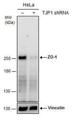
- Experimental details
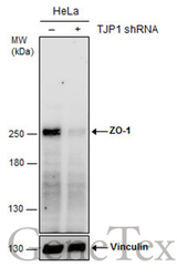
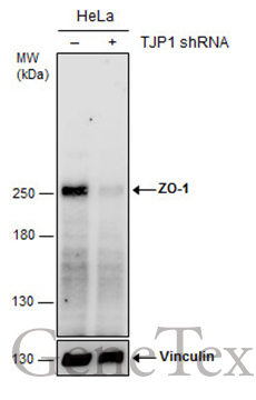
- Non-transfected (¡V) and transfected (+) HeLa whole cell extracts (30 ?g) were separated by 5% SDS-PAGE, and the membrane was blotted with ZO-1 antibody [N1N2], N-term (GTX108613) diluted at 1:1000. The HRP-conjugated anti-rabbit IgG antibody (GTX213110-01) was used to detect the primary antibody.
Supportive validation
- Submitted by
- GeneTex (provider)
- Main image
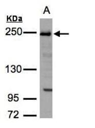
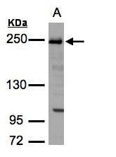
- Experimental details
- Sample(30 ?g whole cell lysate)A:H12995% SDS PAGEGTX108613 diluted at 1:3000The HRP-conjugated anti-rabbit IgG antibody (GTX213110-01) was used to detect the primary antibody.
- Submitted by
- GeneTex (provider)
- Main image
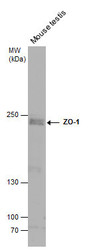
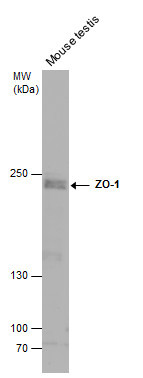
- Experimental details
- Mouse tissue extract (50 ?g) was separated by 5% SDS-PAGE, and the membrane was blotted with ZO-1 antibody [N1N2], N-term (GTX108613) diluted at 1:500. The HRP-conjugated anti-rabbit IgG antibody (GTX213110-01) was used to detect the primary antibody.
- Submitted by
- GeneTex (provider)
- Main image
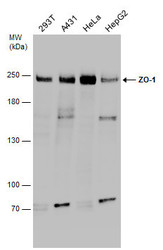
- Experimental details
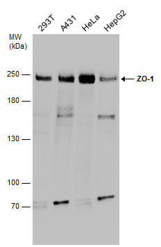
- Various whole cell extracts (30 ?g) were separated by 5% SDS-PAGE, and the membrane was blotted with ZO-1 antibody [N1N2], N-term (GTX108613) diluted at 1:1000.
- Submitted by
- GeneTex (provider)
- Main image

- Experimental details
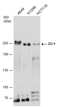
- Various whole cell extracts (30 ?g) were separated by 5% SDS-PAGE, and the membrane was blotted with ZO-1 antibody [N1N2], N-term (GTX108613) diluted at 1:3000.
- Submitted by
- GeneTex (provider)
- Main image
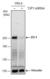
- Experimental details
- Non-transfected (¡V) and transfected (+) HeLa whole cell extracts (30 ?g) were separated by 5% SDS-PAGE, and the membrane was blotted with ZO-1 antibody [N1N2], N-term (GTX108613) diluted at 1:1000. The HRP-conjugated anti-rabbit IgG antibody (GTX213110-01) was used to detect the primary antibody.
Supportive validation
- Submitted by
- GeneTex (provider)
- Main image
- Experimental details
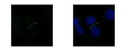
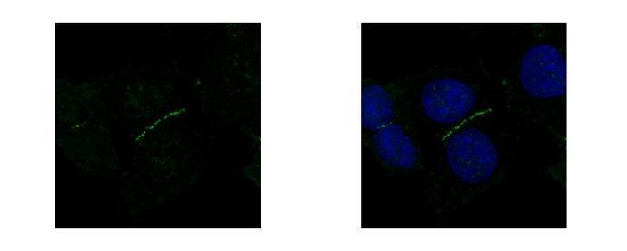
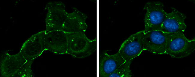
- ZO-1 antibody [N1N2], N-term detects TJP1 protein at junction by confocal immunofluorescent analysis. Sample: A431 cells were fixed in 4% paraformaldehyde at RT for 15 min.Green: TJP1 protein stained by ZO-1 antibody [N1N2], N-term (GTX108613) diluted at 1:500.Blue: Hoechst 33342 staining.[Images captured by Olympus FV10i Confocal Laser Scanning Microscope]
- Submitted by
- GeneTex (provider)
- Main image
- Experimental details
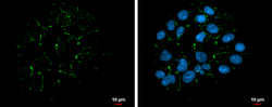
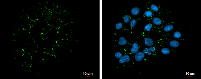
- ZO-1 antibody [N1N2], N-term detects ZO-1 protein at cell membrane by immunofluorescent analysis.Sample: NT2D1 cells were fixed in ice-cold MeOH for 5 min.Green: ZO-1 protein stained by ZO-1 antibody [N1N2], N-term (GTX108613) diluted at 1:200.Blue: Hoechst 33342 staining.Scale bar = 10 £gm.
- Submitted by
- GeneTex (provider)
- Main image

- Experimental details
- ZO-1 antibody [N1N2], N-term detects ZO-1 protein at cell membrane by immunofluorescent analysis.Sample: NT2D1 cells were fixed in ice-cold MeOH for 5 min.Green: ZO-1 stained by ZO-1 antibody [N1N2], N-term (GTX108613) diluted at 1:200.Blue: Hoechst 33342 staining.
Supportive validation
- Submitted by
- GeneTex (provider)
- Main image
- Experimental details
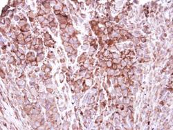
- Immunohistochemical analysis of paraffin-embedded H661 xenograft, using ZO-1(GTX108613) antibody at 1:250 dilution.
- Submitted by
- GeneTex (provider)
- Main image
- Experimental details
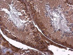
- ZO-1 antibody [N1N2], N-term detects ZO-1 protein at cell membrane and cytoplasm in mouse testis by immunohistochemical analysis. Sample: Paraffin-embedded mouse testis. ZO-1 antibody [N1N2], N-term (GTX108613) diluted at 1:500.

- Submitted by
- GeneTex (provider)
- Main image
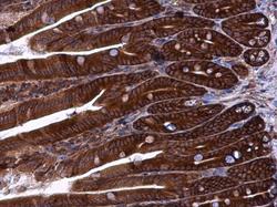
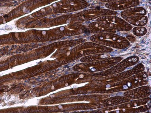
- Experimental details
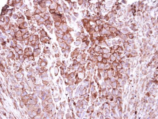
- ZO-1 antibody [N1N2], N-term detects ZO-1 protein at cell membrane and cytoplasm in mouse intestine by immunohistochemical analysis. Sample: Paraffin-embedded mouse intestine. ZO-1 antibody [N1N2], N-term (GTX108613) diluted at 1:500.