 Explore
 Validate
 Learn
Learn Western blot
Antibody data
- Antibody Data
- Antigen structure
- References [9]
- Comments [0]
- Validations
- Western blot [1]
- ELISA [1]
- Immunohistochemistry [1]
Submit
Validation data
Reference
Comment
Report error
- Product number
- AF1819 - Provider product page

- Provider
- R&D Systems
- Product name
- Mouse Klotho Antibody
- Antibody type
- Polyclonal
- Description
- Antigen Affinity-purified. Detects mouse Klotho in direct ELISAs and Western blots. In direct ELISAs and Western blots, less than 5% cross-reactivity with recombinant mouse beta-Klotho is observed.
- Reactivity
- Mouse
- Host
- Goat
- Conjugate
- Unconjugated
- Antigen sequence
BAA25307- Isotype
- IgG
- Vial size
- 100 ug
- Concentration
- LYOPH
- Storage
- Use a manual defrost freezer and avoid repeated freeze-thaw cycles. 12 months from date of receipt, -20 to -70 °C as supplied. 1 month, 2 to 8 °C under sterile conditions after reconstitution. 6 months, -20 to -70 °C under sterile conditions after reconstitution.
Submitted references Secreted Klotho Attenuates Inflammation-Associated Aortic Valve Fibrosis in Senescence-Accelerated Mice P1.
Klotho expression is a prerequisite for proper muscle stem cell function and regeneration of skeletal muscle.
Persistent fibroblast growth factor 23 signalling in the parathyroid glands for secondary hyperparathyroidism in mice with chronic kidney disease.
Antiaging Gene Klotho Attenuates Pancreatic β-Cell Apoptosis in Type 1 Diabetes.
FGF23 suppresses chondrocyte proliferation in the presence of soluble α-Klotho both in vitro and in vivo.
Expression of klotho mRNA and protein in rat brain parenchyma from early postnatal development into adulthood.
Loss of Klotho contributes to kidney injury by derepression of Wnt/β-catenin signaling.
Vitamin D receptor agonists increase klotho and osteopontin while decreasing aortic calcification in mice with chronic kidney disease fed a high phosphate diet.
Circulating αKlotho influences phosphate handling by controlling FGF23 production.
Chen J, Fan J, Wang S, Sun Z
Hypertension (Dallas, Tex. : 1979) 2018 May;71(5):877-885
Hypertension (Dallas, Tex. : 1979) 2018 May;71(5):877-885
Klotho expression is a prerequisite for proper muscle stem cell function and regeneration of skeletal muscle.
Ahrens HE, Huettemeister J, Schmidt M, Kaether C, von Maltzahn J
Skeletal muscle 2018 Jul 4;8(1):20
Skeletal muscle 2018 Jul 4;8(1):20
Persistent fibroblast growth factor 23 signalling in the parathyroid glands for secondary hyperparathyroidism in mice with chronic kidney disease.
Kawakami K, Takeshita A, Furushima K, Miyajima M, Hatamura I, Kuro-O M, Furuta Y, Sakaguchi K
Scientific reports 2017 Jan 17;7:40534
Scientific reports 2017 Jan 17;7:40534
Antiaging Gene Klotho Attenuates Pancreatic β-Cell Apoptosis in Type 1 Diabetes.
Lin Y, Sun Z
Diabetes 2015 Dec;64(12):4298-311
Diabetes 2015 Dec;64(12):4298-311
FGF23 suppresses chondrocyte proliferation in the presence of soluble α-Klotho both in vitro and in vivo.
Kawai M, Kinoshita S, Kimoto A, Hasegawa Y, Miyagawa K, Yamazaki M, Ohata Y, Ozono K, Michigami T
The Journal of biological chemistry 2013 Jan 25;288(4):2414-27
The Journal of biological chemistry 2013 Jan 25;288(4):2414-27
Expression of klotho mRNA and protein in rat brain parenchyma from early postnatal development into adulthood.
Clinton SM, Glover ME, Maltare A, Laszczyk AM, Mehi SJ, Simmons RK, King GD
Brain research 2013 Aug 21;1527:1-14
Brain research 2013 Aug 21;1527:1-14
Loss of Klotho contributes to kidney injury by derepression of Wnt/β-catenin signaling.
Zhou L, Li Y, Zhou D, Tan RJ, Liu Y
Journal of the American Society of Nephrology : JASN 2013 Apr;24(5):771-85
Journal of the American Society of Nephrology : JASN 2013 Apr;24(5):771-85
Vitamin D receptor agonists increase klotho and osteopontin while decreasing aortic calcification in mice with chronic kidney disease fed a high phosphate diet.
Lau WL, Leaf EM, Hu MC, Takeno MM, Kuro-o M, Moe OW, Giachelli CM
Kidney international 2012 Dec;82(12):1261-70
Kidney international 2012 Dec;82(12):1261-70
Circulating αKlotho influences phosphate handling by controlling FGF23 production.
Smith RC, O'Bryan LM, Farrow EG, Summers LJ, Clinkenbeard EL, Roberts JL, Cass TA, Saha J, Broderick C, Ma YL, Zeng QQ, Kharitonenkov A, Wilson JM, Guo Q, Sun H, Allen MR, Burr DB, Breyer MD, White KE
The Journal of clinical investigation 2012 Dec;122(12):4710-5
The Journal of clinical investigation 2012 Dec;122(12):4710-5
No comments: Submit comment
Supportive validation
- Submitted by
- R&D Systems (provider)
- Main image
- Experimental details
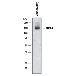
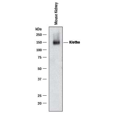
- Detection of Mouse Klotho by Western Blot. Western blot shows lysates of mouse kidney tissue. PVDF membrane was probed with 0.2 µg/mL of Goat Anti-Mouse Klotho Antigen Affinity-purified Polyclonal Antibody (Catalog # AF1819) followed by HRP-conjugated Anti-Goat IgG Secondary Antibody (Catalog # HAF109). A specific band was detected for Klotho at approximately 145 kDa (as indicated). This experiment was conducted under reducing conditions and using Immunoblot Buffer Group 1.
Supportive validation
- Submitted by
- R&D Systems (provider)
- Main image
- Experimental details
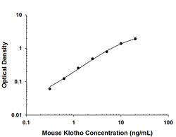
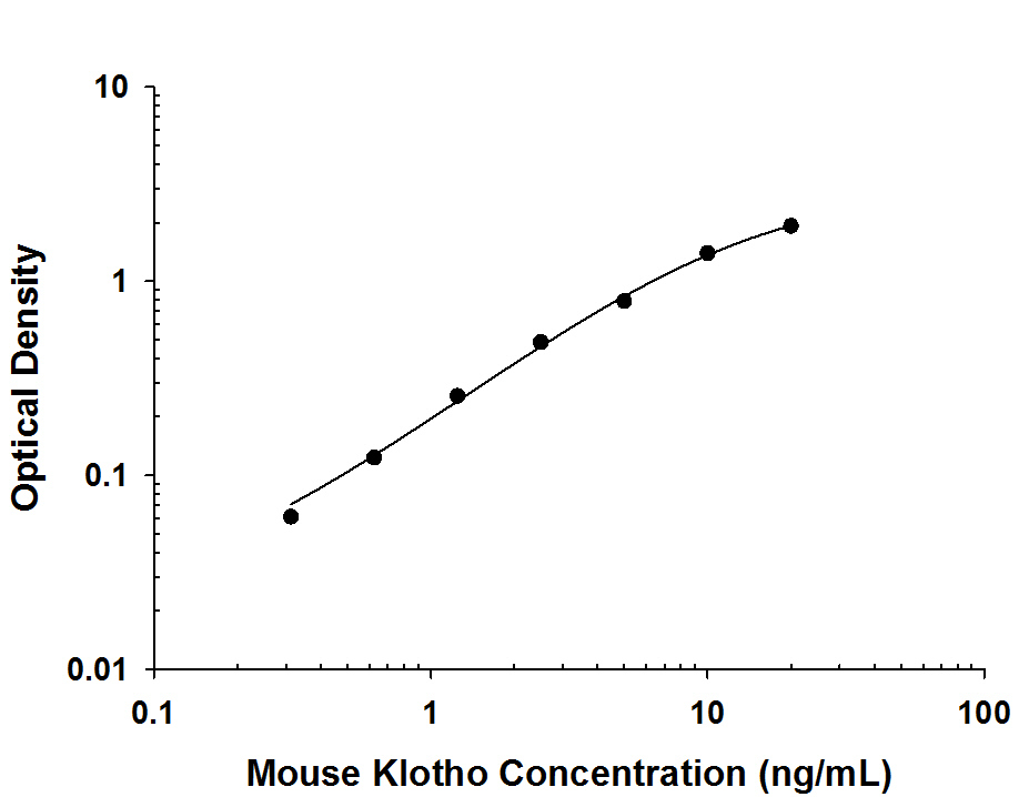
- Mouse Klotho ELISA Standard Curve. Recombinant Mouse Klotho protein was serially diluted 2-fold and captured by Rat Anti-Mouse Klotho Monoclonal Antibody (Catalog # MAB1819) coated on a Clear Polystyrene Microplate (Catalog # DY990). Goat Anti-Mouse Klotho Antigen Affinity-purified Polyclonal Antibody (Catalog # AF1819) was biotinylated and incubated with the protein captured on the plate. Detection of the standard curve was achieved by incubating Streptavidin-HRP (Catalog # DY998) followed by Substrate Solution (Catalog # DY999) and stopping the enzymatic reaction with Stop Solution (Catalog # DY994).
Supportive validation
- Submitted by
- R&D Systems (provider)
- Main image
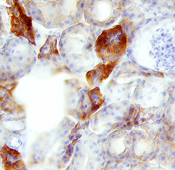
- Experimental details
- Klotho in Mouse Kidney. Klotho was detected in perfusion fixed frozen sections of mouse kidney using 1.7 µg/mL Goat Anti-Mouse Klotho Antigen Affinity-purified Polyclonal Antibody (Catalog # AF1819) overnight at 4 °C. Tissue was stained with the Anti-Goat HRP-DAB Cell & Tissue Staining Kit (brown; Catalog # CTS008) and counterstained with hematoxylin (blue). View our protocol for Chromogenic IHC Staining of Frozen Tissue Sections.
