 Explore
 Validate
 Learn
Learn Western blot
Antibody data
- Antibody Data
- Antigen structure
- References [0]
- Comments [0]
- Validations
- Western blot [6]
- Immunocytochemistry [1]
- Immunohistochemistry [1]
Submit
Validation data
Reference
Comment
Report error
- Product number
- PA5-30221 - Provider product page

- Provider
- Invitrogen Antibodies
- Product name
- PCK2 Polyclonal Antibody
- Antibody type
- Polyclonal
- Antigen
- Recombinant protein fragment
- Description
- Recommended positive controls: 293T, HeLa, HepG2, Neuro2A, GL261, PC-12.
- Concentration
- 1 mg/mL
No comments: Submit comment
Supportive validation
- Submitted by
- Invitrogen Antibodies (provider)
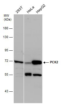
- Main image

- Experimental details
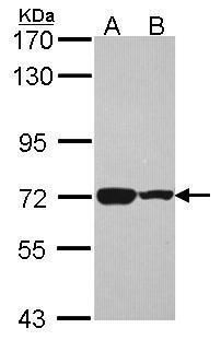
- Western blot analysis of PCK2 using 30 µg of A) HepG2 and B) HCT116 lysate. Samples were loaded onto a 7.5% SDS-PAGE gel and probed with a PCK2 polyclonal antibody (Product # PA5-30221) at a dilution of 1:1000.
- Submitted by
- Invitrogen Antibodies (provider)
- Main image
- Experimental details
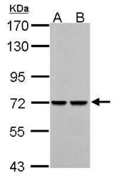
- PCK2 Polyclonal Antibody detects PCK2 protein by western blot analysis. A. 30 µg Neuro2A whole cell lysate/extract. B. 30 µg GL261 whole cell lysate/extract.7.5% SDS-PAGE. PCK2 Polyclonal Antibody (Product # PA5-30221) dilution: 1:1,000. The HRP-conjugated anti-rabbit IgG antibody was used to detect the primary antibody.

- Submitted by
- Invitrogen Antibodies (provider)
- Main image
- Experimental details
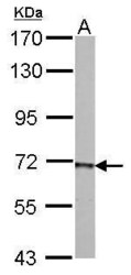
- PCK2 Polyclonal Antibody detects PCK2 protein by western blot analysis. A. 30 µg PC-12 whole cell lysate/extract.7.5% SDS-PAGE. PCK2 Polyclonal Antibody (Product # PA5-30221) dilution: 1:1,000. The HRP-conjugated anti-rabbit IgG antibody was used to detect the primary antibody.
- Submitted by
- Invitrogen Antibodies (provider)
- Main image

- Experimental details
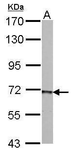
- Western Blot analysis of PEPCK was performed by separating 30 µg of various whole cell extracts by 7.5% SDS-PAGE. Proteins were transferred to a membrane and probed with a PEPCK Polyclonal Antibody (Product # PA5-30221) at a dilution of 1:500 and a HRP-conjugated anti-rabbit IgG secondary antibody.
- Submitted by
- Invitrogen Antibodies (provider)
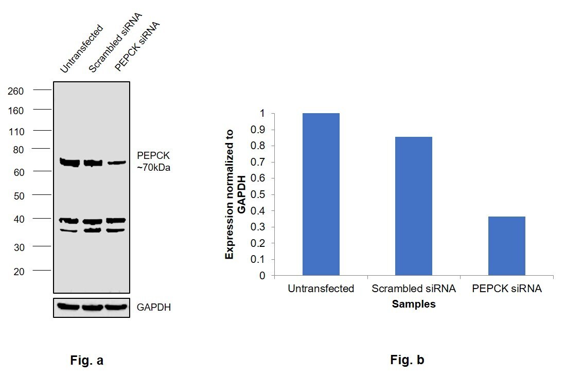
- Main image

- Experimental details
- Knockdown of PEPCK was achieved by transfecting A549 cells with PEPCK specific siRNAs (Silencer® select Product # Retrieving data. Wait a few seconds and try to cut or copy again.). Western blot analysis (Fig. a) was performed using Membrane enriched extracts from the PEPCK knockdown cells (lane 3), non-targeting scrambled siRNA transfected cells (lane 2) and untransfected cells (lane 1). The blot was probed with PEPCK Polyclonal Antibody (Product # PA5-30221, 1:1000 ) and Goat anti-Rabbit IgG (H+L) Superclonal™ Recombinant Secondary Antibody, HRP (Product # A27036, 1:4000). Densitometric analysis of this western blot is shown in histogram (Fig. b). Decrease in signal upon siRNA mediated knock down confirms that antibody is specific to PEPCK. Two non specific bands around 36 to 40kDa were observed.
- Submitted by
- Invitrogen Antibodies (provider)
- Main image
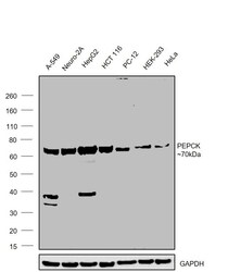
- Experimental details
- Western blot was performed using Anti-PEPCK Polyclonal Antibody (Product # PA5-30221) and a ~70kDa band corresponding to PEPCK was observed across cell lines tested. Membrane enriched extracts (30 µg lysate) of A549 (Lane 1), Neuro-2a (Lane 2), Hep G2 (Lane 3), HCT 116 (Lane 4), PC-12 (Lane 5), HEK-293 (Lane 6), HeLa (Lane 7) were electrophoresed using NuPAGE™ 4-12% Bis-Tris Protein Gel (Product # NP0321BOX). Resolved proteins were then transferred onto a Nitrocellulose membrane (Product # LC2001) by iBlot® 2 Dry Blotting System (Product # IB21001). The blot was probed with the primary antibody (1:1000) and detected by chemiluminescence with Goat anti-Rabbit IgG (H+L) Superclonal™ Recombinant Secondary Antibody, HRP (Product # A27036,1:4000) using the iBright FL 1000 (Product # A32752). Chemiluminescent detection was performed using Novex® ECL Chemiluminescent Substrate Reagent Kit (Product # WP20005).

Supportive validation
- Submitted by
- Invitrogen Antibodies (provider)
- Main image
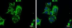
- Experimental details
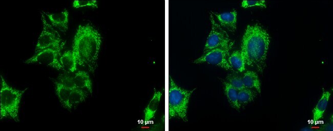
- Immunocytochemistry-Immunofluorescence analysis of PEPCK was performed in HepG2 cells fixed in MeOH for 5 min. Green: PEPCK Polyclonal Antibody (Product # PA5-30221) diluted at 1:500. Blue: Hoechst 33342 staining. Scale bar = 10 µm.
Supportive validation
- Submitted by
- Invitrogen Antibodies (provider)
- Main image
- Experimental details
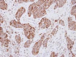
- Immunohistochemical analysis of paraffin-embedded human breast cancer, using PCK2 (Product # PA5-30221) antibody at 1:250 dilution. Antigen Retrieval: EDTA based buffer, pH 8.0, 15 min.
