Explore
 Validate
 Learn
Learn Western blot
Antibody data
- Antibody Data
- Antigen structure
- References [0]
- Comments [0]
- Validations
- Western blot [3]
- Immunocytochemistry [3]
- Immunohistochemistry [3]
- Other assay [1]
Submit
Validation data
Reference
Comment
Report error
- Product number
- PA5-22102 - Provider product page

- Provider
- Invitrogen Antibodies
- Product name
- GLDC Polyclonal Antibody
- Antibody type
- Polyclonal
- Antigen
- Recombinant protein fragment
- Description
- Recommended positive controls: HePG2.
- Concentration
- 0.46 mg/mL
No comments: Submit comment
Supportive validation
- Submitted by
- Invitrogen Antibodies (provider)
- Main image
- Experimental details
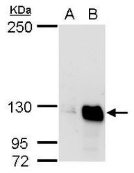
- Western blot analysis of glycine dehydrogenase (decarboxylating) precursor using A) 30 µg HeLa whole cell lysate and B) 30 µg HepG2 whole cell lysate. Samples were loaded onto a 5% SDS-PAGE gel and probed with a glycine dehydrogenase (decarboxylating) precursor polyclonal antibody (Product # PA5-22102) at a dilution of 1:500.

- Submitted by
- Invitrogen Antibodies (provider)
- Main image

- Experimental details
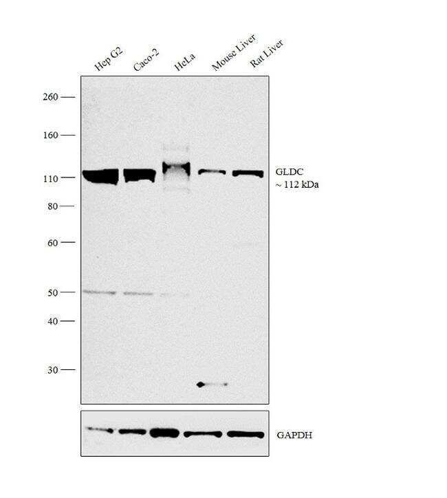
- Western blot analysis was performed on membrane enriched extracts (30 µg lysate) of HepG2 (Lane 1), Caco-2 (Lane 2), HeLa (Lane 3), tissue extract of Mouse Liver (Lane 4) and Rat Liver (Lane 5). The blot was probed with Anti-GLDC Polyclonal Antibody (Product # PA5-22102, 1:500 dilution) and detected by chemiluminescence using Goat anti-Rabbit IgG (H+L) Superclonal™ Secondary Antibody, HRP conjugate (Product # A27036, 0.25 µg/ml, 1:4000 dilution). A 112 kDa band corresponding to GLDC was observed across the cell lines and tissues tested.
- Submitted by
- Invitrogen Antibodies (provider)
- Main image
- Experimental details
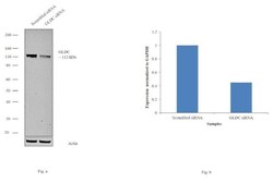
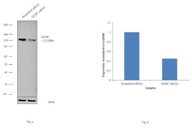
- Knockdown of GLDC was achieved by transfecting HepG2 cells with GLDC specific siRNAs (Silencer® select Product # s223750, s5805). Western blot analysis (Fig. a) was performed using membrane enriched extracts from the GLDC knockdown cells (lane 3), non-specific scrambled siRNA transfected cells (lane 2) and untransfected cells (lane 1). The blot was probed with GLDC Polyclonal Antibody (Product # PA5-22102, 1:2000 dilution) and Goat anti-Rabbit IgG (H+L) Superclonal™ Secondary Antibody, HRP conjugate (Product # A27036, 0.25µg/ml, 1:4000 dilution). Densitometric analysis of this western blot is shown in histogram (Fig. b). Decrease in signal upon siRNA mediated knock down confirms that antibody is specific to GLDC.
Supportive validation
- Submitted by
- Invitrogen Antibodies (provider)
- Main image
- Experimental details
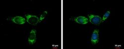
- Immunofluorescent analysis of glycine dehydrogenase (decarboxylating) precursor showing staining in the mitochondria of HepG2 cells. HepG2 cells were fixed in ice-cold MeOH for 5 min and stained using a glycine dehydrogenase (decarboxylating) precursor polyclonal antibody (Product # PA5-22102) diluted at 1:500. Blue: Hoechst 33342 staining.
- Submitted by
- Invitrogen Antibodies (provider)
- Main image
- Experimental details
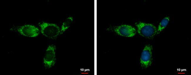
- Immunocytochemistry-Immunofluorescence analysis of GLDC was performed in Hep G2 cells fixed in ice-cold MeOH for 5 min. Green: GLDC Polyclonal Antibody (Product # PA5-22102) diluted at 1:400. Blue: Hoechst 33342 staining.

- Submitted by
- Invitrogen Antibodies (provider)
- Main image
- Experimental details
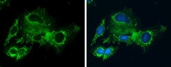
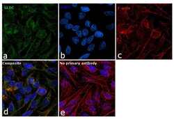
- Immunofluorescence analysis of GLDC was performed using 70% confluent log phase HeLa cells. The cells were fixed with ice-cold acetone for 5 minutes and blocked with 1% BSA for 1 hour at room temperature. The cells were labeled with GLDC Polyclonal Antibody (Product # PA5-22102) at 1:100 dilution in 0.1% BSA, incubated at 4 degree celsius overnight and then labeled with Goat anti-Rabbit IgG (H+L) Superclonal™ Secondary Antibody, Alexa Fluor® 488 conjugate (Product # A27034) at a dilution of 1:2000 for 45 minutes at room temperature (Panel a: green).Nuclei (Panel b: blue) were stained with SlowFade® Gold Antifade Mountant with DAPI (Product # S36938). F-actin (Panel c: red) was stained with Rhodamine Phalloidin (Product # R415, 1:300). Panel d represents the merged image showing mitochondrial and nuclear localization. Panel e represents control cells with no primary antibody to assess background. The images were captured at 60X magnification.
Supportive validation
- Submitted by
- Invitrogen Antibodies (provider)
- Main image

- Experimental details
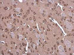
- Immunohistochemistry (Paraffin) analysis of GLDC was performed in paraffin-embedded mouse liver tissue using GLDC Polyclonal Antibody (Product # PA5-22102) at a dilution of 1:500.

- Submitted by
- Invitrogen Antibodies (provider)
- Main image
- Experimental details
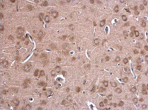
- Immunohistochemistry (Paraffin) analysis of GLDC was performed in paraffin-embedded mouse brain tissue using GLDC Polyclonal Antibody (Product # PA5-22102) at a dilution of 1:500.
- Submitted by
- Invitrogen Antibodies (provider)
- Main image

- Experimental details
- Immunohistochemistry (Paraffin) analysis of GLDC was performed in paraffin-embedded rat brain tissue using GLDC Polyclonal Antibody (Product # PA5-22102) at a dilution of 1:500.
Supportive validation
- Submitted by
- Invitrogen Antibodies (provider)
- Main image
- Experimental details
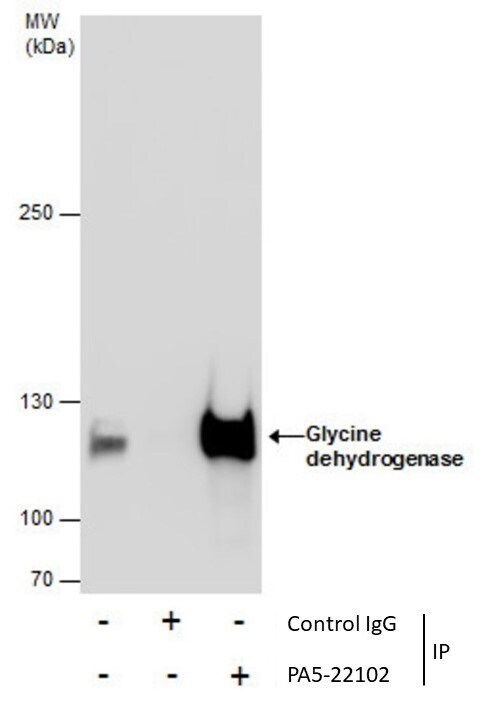
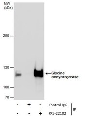
- Immunoprecipitation of Glycine dehydrogenase was performed in HepG2 whole cell extracts using 5 µg of GLDC Polyclonal Antibody (Product # PA5-22102). Samples were transferred to a membrane and probed with GLDC Polyclonal Antibody as a primary antibody and an HRP-conjugated anti-Rabbit IgG was used as a secondary antibody.