Explore
 Validate
 Learn
Learn Western blot
 Immunohistochemistry
Antibody data
- Antibody Data
- Antigen structure
- References [0]
- Comments [0]
- Validations
- Immunohistochemistry [1]
- Flow cytometry [2]
Submit
Validation data
Reference
Comment
Report error
- Product number
- NBP2-34752-0.1 mg - Provider product page

- Provider
- Novus Biologicals
- Product name
- Mouse Monoclonal Cytokeratin 10 Antibody
- Antibody type
- Monoclonal
- Description
- Protein G purified. This MAb recognizes a protein of 56.5kDa, identified as cytokeratin 10 (CK10). CK10 is expressed in all suprabasal layers of the epidermis. In the epidermis, expression of CK10 strictly parallels the extent of differentiation
- Reactivity
- Human, Mouse
- Host
- Mouse
- Isotype
- IgG
- Vial size
- 0.1 mg
- Concentration
- 1.0 mg/ml
- Storage
- Store at 4C short term. Aliquot and store at -20C long term. Avoid freeze-thaw cycles.
No comments: Submit comment
Supportive validation
- Submitted by
- Novus Biologicals (provider)
- Main image

- Experimental details
- Immunohistochemistry-Paraffin: Cytokeratin 10 Antibody (SPM261) - Azide and BSA Free [NBP2-34752] - Formalin-fixed, paraffin-embedded human Bladder Carcinoma stained with Cytokeratin 10 Antibody (SPM261).

Supportive validation
- Submitted by
- Novus Biologicals (provider)
- Main image

- Experimental details
- Flow Cytometry: Cytokeratin 10 Antibody (SPM261) - Azide and BSA Free [NBP2-34752] - An intracellular stain was performed on A431 cells with Cytokeratin 10 [SPM261] Antibody NBP2-34752B (blue) and a matched isotype control (orange). Both antibodies were conjugated to Biotin. Cells were fixed with 4% PFA and then permeabilized with 0.1% saponin. Cells were incubated in an antibody dilution of 2.5 ug/mL for 30 minutes at room temperature, followed by Streptavidin - R-Phycoerythrin Protein (2012-1000, Novus Biologicals).

- Submitted by
- Novus Biologicals (provider)
- Main image

- Experimental details
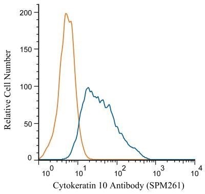
- Flow Cytometry: Cytokeratin 10 Antibody (SPM261) - Azide and BSA Free [NBP2-34752] - Analysis using Alexa Fluor (R) 647 conjugate of NBP2-32962. An intracellular stain was performed on HeLa cells with Cytokeratin 10 antibody (SPM261) NBP2-34752 (blue) and a matched isotype control NBP2-27287 (orange). Cells were fixed with 4% PFA and then permeablized with 0.1% saponin. 1 microgram of antibody was added to 100 uL of staining buffer and cells were incubated for 30 minutes at room temperature. Both antibodies were conjugated to Alexa Fluor 647.