Explore
 Validate
 Learn
Learn Western blot
 Immunocytochemistry
 Immunohistochemistry
Antibody data
- Antibody Data
- Antigen structure
- References [0]
- Comments [0]
- Validations
- Immunohistochemistry [2]
- Other assay [1]
Submit
Validation data
Reference
Comment
Report error
- Product number
- STJA0003676 - Provider product page

- Provider
- St John's Laboratory
- Product name
- Anti-MAP2 antibody (STJA0003676)
- Antibody type
- Polyclonal
- Description
- Chicken polyclonal antibody anti-MAP2 is suitable for use in Western Blot and Immunofluorescence research applications.
- Reactivity
- Human, Mouse, Rat, Bovine
- Host
- Chicken/Avian
- Conjugate
- Unconjugated
- Antigen sequence
NA- Epitope
- NA
- Antibody clone number
- NA
- Vial size
- NA
- Concentration
- NA
- Storage
- Store at-20°C, and avoid repeated freeze-thaw cycles.
- Handling
- NA
No comments: Submit comment
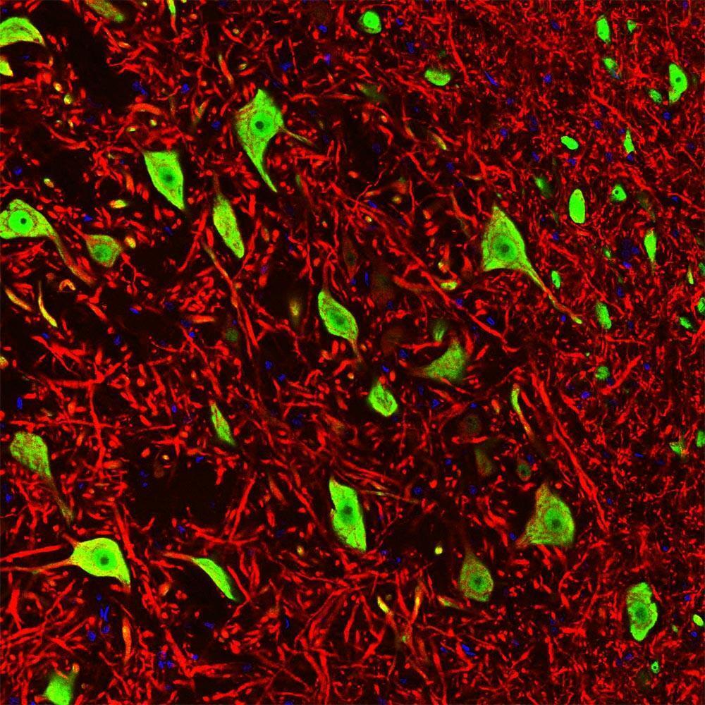
Supportive validation
Supportive validation
- Submitted by
- St John's Laboratory (provider)
- Main image
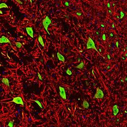
- Experimental details
- Immunofluorescence of a section of rat brain stem showing colabeled with Anti-MAP2 (cat. STJA0003676, red, 1:5000) and Anti-FOX3 ( cat. STJA0003841, green, 1:1000). The Anti-FOX3 specifically labels the nuclei and the proximal cytoplasm of neuronal cells while the Anti-MAP2 labels dendrites and overlaps with FOX3 labeling the perikarya of neurons. The blue is DAPI staining of nuclear DNA.
- Sample type
- NA
- Validation comment
- NA
- Primary Ab dilution
- NA
- Other comments
- NA
- Secondary Ab
- NA
- Secondary Ab dilution
- NA
- Protocol
- NA
Supportive validation
- Submitted by
- St John's Laboratory (provider)
- Main image
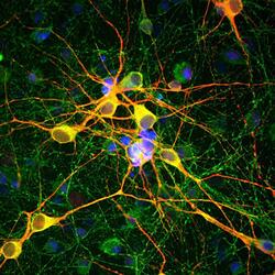
- Experimental details
- Immunofluorescence of cortical neuron-glial cell culture from E20 rat labeled with Anti-MAP2 (cat. STJA0003676, red, 1:10,000) , and Anti-Tau (green). The blue is DAPI staining of nuclear DNA. The anti-MAP2 antibody stains dendrites and perikarya of neurons, while the anti-TAU antibody labels neuronal perikarya, dendrites and also axonal process. As a result perikarya and dendrites appears orange-yellow, since they contain both MAP2 and tau, while axons are green.
- Sample type
- NA
- Validation comment
- NA
- Primary Ab dilution
- NA
- Other comments
- NA
- Secondary Ab
- NA
- Secondary Ab dilution
- NA
- Protocol
- NA

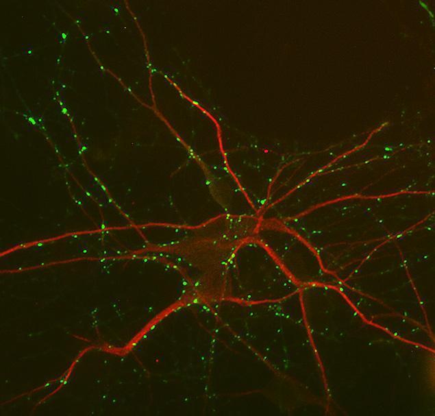
Supportive validation
- Submitted by
- St John's Laboratory (provider)
- Main image
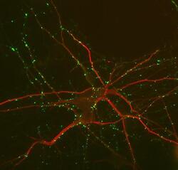
- Experimental details
- Immunolabeling of mouse hippocampal cultures labeled with anti-MAP2 (cat. STJA0003676, 1:1000, red) and anti-synapsin (cat. STJA0003781, 1:1000, green).
- Sample type
- NA
- Validation comment
- NA
- Primary Ab dilution
- NA
- Other comments
- NA
- Secondary Ab
- NA
- Secondary Ab dilution
- NA
- Protocol
- NA