Explore
 Validate
 Learn
Learn Western blot
Antibody data
- Antibody Data
- Antigen structure
- References [0]
- Comments [0]
- Validations
- Western blot [1]
- ELISA [1]
- Immunocytochemistry [2]
- Immunohistochemistry [1]
Submit
Validation data
Reference
Comment
Report error
- Product number
- 101-M09 - Provider product page

- Provider
- ReliaTech GmbH
- Product name
- Anti-human EMILIN1
- Antibody type
- Monoclonal
- Description
- antibody Protein-G purified from hybridoma supernatant
- Reactivity
- Human
- Host
- Mouse
- Antibody clone number
- (#1H2/G8)
- Storage
- The lyophilized antibody is stable for at least 2 years at -20°C. After sterile reconstitution the antibody is stable at 2-8°C for up to 6 months. Frozen aliquots are stable for at least 6 months when stored at -20°C. Addition of a carrier protein or 50% glycerol is recommended for frozen aliquots.
- Handling
- Centrifuge vial prior to opening. Reconstitute in sterile water to a concentration of 0.1-1.0 mg/ml.
No comments: Submit comment
Supportive validation
- Submitted by
- ReliaTech GmbH (provider)
- Main image

- Experimental details
- Western blot analysis with anti-human EMILIN1 #1H2/G8 antibody [Cat# 101-M09] on recombinant human EMILIN1 purified from HEK293 transfected cells.

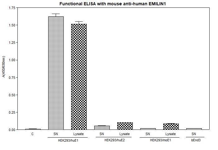
Supportive validation
- Submitted by
- ReliaTech GmbH (provider)
- Main image

- Experimental details
- Functional ELISA: Supernatant (200μl) and total cell lysate (100μl, 1:2 diluted) of HEK293 cells expressing the different EMILIN proteins were coated to a 96-well plate and incubated at 37°C for >1 hour. After blocking and several wash steps the #1H2/G8 [Cat# 101-M09] antibody was added (2μg/ml) and incubated for another 1 hour. Detection was performed with an anti-mouse secondary antibody.
Supportive validation
- Submitted by
- ReliaTech GmbH (provider)
- Main image

- Experimental details
- Immunofluorescence staining of normal cultured cells (skin human fibroblasts) (panel a, left) and of cryostat tissue section (undifferentiated soft tissue sarcoma) (panel b, right) with #1H2/G8 [Cat# 101-M09]) (green staining). Nuclei are in blue. Dotted lines indicate the border between “normal” (lower corner) and “tumor” tissue.

- Submitted by
- ReliaTech GmbH (provider)
- Main image

- Experimental details
- Immunofluorescence staining of human EMILIN1 in HEK293 cells expressing human EMILIN1 with the monoclonal mouse anti-human EMILIN1 antibody #1H2/G8 [Cat# 101-M09]. Conjugated secondary antibody: goat anti-mouse ALEXA Flour 488 (1:600) [Dianova].

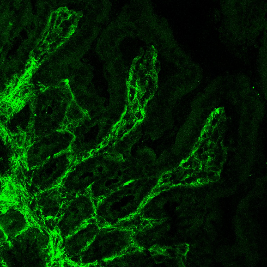
Supportive validation
- Submitted by
- ReliaTech GmbH (provider)
- Main image

- Experimental details
- Immunofluorescence staining of human intestine cryostat sections with the monoclonal mouse anti-human EMILIN1 antibody #1H2/G8 [Cat# 101-M09].
- Sample type
- human intestine cryostat section