PA5-20812
antibody from Invitrogen Antibodies
Targeting: AGTR1
AG2S, AGTR1A, AGTR1B, AT1, AT1B, AT2R1, AT2R1A, AT2R1B, HAT1R
Antibody data
- Antibody Data
- Antigen structure
- References [6]
- Comments [0]
- Validations
- Western blot [2]
- Immunocytochemistry [2]
- Immunohistochemistry [2]
- Other assay [6]
Submit
Validation data
Reference
Comment
Report error
- Product number
- PA5-20812 - Provider product page

- Provider
- Invitrogen Antibodies
- Product name
- AGTR1 Polyclonal Antibody
- Antibody type
- Polyclonal
- Antigen
- Synthetic peptide
- Description
- A suggested positive control is mouse kidney tissue lysate. PA5-20812 can be used with blocking peptide PEP-0926.
- Reactivity
- Human, Mouse, Rat
- Host
- Rabbit
- Isotype
- IgG
- Vial size
- 100 µg
- Concentration
- 1 mg/mL
- Storage
- Maintain refrigerated at 2-8°C for up to 3 months. For long term storage store at -20°C
Submitted references Myogenic Vasoconstriction Requires Canonical G(q/11) Signaling of the Angiotensin II Type 1 Receptor.
Effect of Angiotensin-Converting-Enzyme Inhibitor and Angiotensin II Receptor Antagonist Treatment on ACE2 Expression and SARS-CoV-2 Replication in Primary Airway Epithelial Cells.
Spent Hen Muscle Protein-Derived RAS Regulating Peptides Show Antioxidant Activity in Vascular Cells.
Effect of (-)-epicatechin on the modulation of progression markers of chronic renal damage in a 5/6 nephrectomy experimental model.
Data supporting that adipose-derived mesenchymal stem/stromal cells express angiotensin II receptors in situ and in vitro.
Differential expression and DNA methylation of angiotensin type 1A receptors in vascular tissues during genetic hypertension development.
Cui Y, Kassmann M, Nickel S, Zhang C, Alenina N, Anistan YM, Schleifenbaum J, Bader M, Welsh DG, Huang Y, Gollasch M
Journal of the American Heart Association 2022 Feb 15;11(4):e022070
Journal of the American Heart Association 2022 Feb 15;11(4):e022070
Effect of Angiotensin-Converting-Enzyme Inhibitor and Angiotensin II Receptor Antagonist Treatment on ACE2 Expression and SARS-CoV-2 Replication in Primary Airway Epithelial Cells.
Okoloko O, Vanderwall ER, Rich LM, White MP, Reeves SR, Harrington WE, Barrow KA, Debley JS
Frontiers in pharmacology 2021;12:765951
Frontiers in pharmacology 2021;12:765951
Spent Hen Muscle Protein-Derived RAS Regulating Peptides Show Antioxidant Activity in Vascular Cells.
Fan H, Bhullar KS, Wu J
Antioxidants (Basel, Switzerland) 2021 Feb 15;10(2)
Antioxidants (Basel, Switzerland) 2021 Feb 15;10(2)
Effect of (-)-epicatechin on the modulation of progression markers of chronic renal damage in a 5/6 nephrectomy experimental model.
Montes-Rivera J, Arellano-Mendoza M, Nájera N, Del Valle-Mondragón L, Villarreal F, Rubio-Gayosso I, Perez-Duran J, Meaney E, Ceballos G
Heliyon 2019 Apr;5(4):e01512
Heliyon 2019 Apr;5(4):e01512
Data supporting that adipose-derived mesenchymal stem/stromal cells express angiotensin II receptors in situ and in vitro.
Ageeva LV, Sysoeva VY, Tyurin-Kuzmin PA, Sharonov GV, Dyikanov DT, Stambolsky DV, Kalinina NI
Data in brief 2018 Feb;16:327-333
Data in brief 2018 Feb;16:327-333
Differential expression and DNA methylation of angiotensin type 1A receptors in vascular tissues during genetic hypertension development.
Pei F, Wang X, Yue R, Chen C, Huang J, Huang J, Li X, Zeng C
Molecular and cellular biochemistry 2015 Apr;402(1-2):1-8
Molecular and cellular biochemistry 2015 Apr;402(1-2):1-8
No comments: Submit comment
Supportive validation
- Submitted by
- Invitrogen Antibodies (provider)
- Main image

- Experimental details
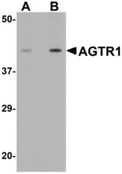
- Western blot analysis of mouse kidney tissue lysate using a AGTR1 polyclonal antibody (Product # PA5-20812) at (A) 1 and (B) 2 µg/mL.
- Submitted by
- Invitrogen Antibodies (provider)
- Main image

- Experimental details
- Western Blot analysis of AGTR1 in mouse kidney tissue lysate with AGTR1 Polyclonal Antibody (Product # PA5-20812) at (A) 1 and (B) 2 µg/mL.
Supportive validation
- Submitted by
- Invitrogen Antibodies (provider)
- Main image

- Experimental details
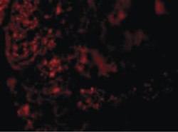
- Immunofluorescent analysis of mouse kidney cells using a AGTR1 polyclonal antibody (Product # PA5-20812) at a 20 µg/mL dilution.
- Submitted by
- Invitrogen Antibodies (provider)
- Main image

- Experimental details
- Immunofluorescence of AGTR1 in Mouse Kidney cells with AGTR1 Polyclonal Antibody (Product # PA5-20812) at 20 µg/mL.
Supportive validation
- Submitted by
- Invitrogen Antibodies (provider)
- Main image

- Experimental details
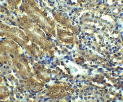
- Immunohistochemistry of AGTR1 in mouse kidney tissue with AGTR1 Polyclonal Antibody (Product # PA5-20812) at 2.5 µg/mL.
- Submitted by
- Invitrogen Antibodies (provider)
- Main image

- Experimental details
- Immunohistochemistry of AGTR1 in mouse kidney tissue with AGTR1 Polyclonal Antibody (Product # PA5-20812) at 2.5 µg/mL.
Supportive validation
- Submitted by
- Invitrogen Antibodies (provider)
- Main image

- Experimental details
- NULL
- Submitted by
- Invitrogen Antibodies (provider)
- Main image

- Experimental details
- NULL
- Submitted by
- Invitrogen Antibodies (provider)
- Main image

- Experimental details
- NULL
- Submitted by
- Invitrogen Antibodies (provider)
- Main image

- Experimental details
- NULL
- Submitted by
- Invitrogen Antibodies (provider)
- Main image

- Experimental details
- Figure 5 Effect of VRP, LKY, VRY, and V-F on the expressions of AT 1 R ( A ) and MasR ( B ) in VSMCs. Cells were treated with peptides (50 muM) for 24 h. Protein bands were quantified by densitometry and normalized to alpha-tubulin. Data were expressed as means +- SEM of 4-5 independent experiments, normalized to the untreated group (Untr); ns: not significant ( p > 0.05); * p < 0.05, as compared to the untreated group.
- Submitted by
- Invitrogen Antibodies (provider)
- Main image

- Experimental details
- FIGURE 7 Immunofluorescence staining for ATR1 and ACE2 in primary differentiated airway epithelial cell cultures infected with SARS-CoV2 and treated with either captopril or losartan. Cultures were infected with SARS-CoV-2 for 96 h and a subset were treated with captopril (1 uM) or losartan (2 uM) throughout the 96-h infection. Depicted are representative confocal images obtained through the apical plane of differentiated airway epithelial cultures; ATR1 staining is shown in green (A) , ACE2 staining is shown in red (B) , and merged images including DAPI staining (blue) are shown in the (C) . No significant differences in staining pattern were observed between the groups (63x objective; Scale bars = 50 um).
 Explore
 Validate
 Learn
Learn Western blot