Explore
 Validate
 Learn
Learn Western blot
 ELISA
ELISAAntibody data
- Antibody Data
- Antigen structure
- References [0]
- Comments [0]
- Validations
- Western blot [1]
Submit
Validation data
Reference
Comment
Report error
- Product number
- A01756-2 - Provider product page

- Provider
- Boster Biological Technology
- Product name
- Anti-CD2AP Antibody Picoband™
- Antibody type
- Polyclonal
- Description
- Polyclonal antibody for CD2AP detection. Host: Rabbit.Size: 100μg/vial. Tested applications: WB, IHC-P, ICC/IF, FCM, Direct ELISA. Reactive species: Human;Mouse;Rat. CD2AP information: Subcellular Localization: Cytoplasm, cytoskeleton . Cell projection, ruffle . Colocalizes with F-actin and BCAR1/p130Cas in membrane ruffles. Located at podocyte slit diaphragm between podocyte foot processes (By similarity). During late anaphase and telophase, concentrates in the vicinity of the midzone microtubules and in the midbody in late telophase; Tissue Specificity: Widely expressed in fetal and adult tissues.
- Reactivity
- Human, Mouse, Rat
- Host
- Rabbit
- Vial size
- 100μg/vial
- Concentration
- 0.5-1mg/ml, actual concentration vary by lot. Use suggested dilution ratio to decide dilution procedure.
- Storage
- At -20°C for one year. After reconstitution, at 4°C for one month. It can also be aliquoted and stored frozen at -20°C for a longer time. Avoid repeated freezing and thawing.
- Handling
- Add 0.2ml of distilled water will yield a concentration of 500ug/ml.
No comments: Submit comment
Supportive validation
- Submitted by
- Boster Biological Technology (provider)
- Main image

- Experimental details
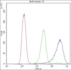
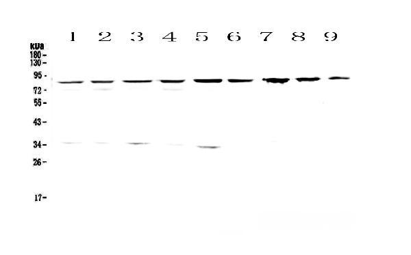
- Western blot analysis of CD2AP using anti-CD2AP antibody (A01756-2). Electrophoresis was performed on a 5-20% SDS-PAGE gel at 70V (Stacking gel) / 90V (Resolving gel) for 2-3 hours. The sample well of each lane was loaded with 50ug of sample under reducing conditions. Lane 1: human Hela whole cell lysates,Lane 2: human 293T whole cell lysates,Lane 3: human MCF-7 whole cell lysates,Lane 4: human COLO-320 whole cell lysates,Lane 5: human 22RV1 whole cell lysates,Lane 6: human SK-OV-3 whole cell lysates,Lane 7: rat stomach tissue lysates,Lane 8: rat liver tissue lysates,Lane 9: mouse liver tissue lysates. After Electrophoresis, proteins were transferred to a Nitrocellulose membrane at 150mA for 50-90 minutes. Blocked the membrane with 5% Non-fat Milk/ TBS for 1.5 hour at RT. The membrane was incubated with rabbit anti-CD2AP antigen affinity purified polyclonal antibody (Catalog # A01756-2) at 0.5 μg/mL overnight at 4°C, then washed with TBS-0.1%Tween 3 times with 5 minutes each and probed with a goat anti-rabbit IgG-HRP secondary antibody at a dilution of 1:10000 for 1.5 hour at RT. The signal is developed using an Enhanced Chemiluminescent detection (ECL) kit (Catalog # EK1002) with Tanon 5200 system. A specific band was detected for CD2AP at approximately 85KD. The expected band size for CD2AP is at 71KD.
- Additional image