Explore
 Validate
 Learn
Learn Western blot
Antibody data
- Antibody Data
- Antigen structure
- References [0]
- Comments [0]
- Validations
- Western blot [1]
- Immunohistochemistry [2]
Submit
Validation data
Reference
Comment
Report error
- Product number
- TA328866 - Provider product page

- Provider
- OriGene
- Product name
- Rabbit Polyclonal Anti-Kisspeptins Receptor (extracellular)
- Antibody type
- Polyclonal
- Description
- Rabbit Polyclonal Anti-Kisspeptins Receptor (extracellular)
- Host
- Rabbit
- Conjugate
- Unconjugated
- Epitope
- KISS1R
- Antibody clone number
- NULL
- Vial size
- 200 µl
- Concentration
- NULL
No comments: Submit comment
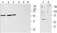
Supportive validation
- Submitted by
- OriGene (provider)
- Main image

- Experimental details
- Western blot analysis of jurkat (lanes 1 and 4), HL-60 (lanes 2 and 5), MCF-7 (lanes 3 and 6) and rat brain (lanes 7 and 8) lysates: 1, 2, 3, 7. Anti-Kisspeptin Receptor (extracellular) antibody, (1:500). 4, 5, 6, 8. Anti-Kisspeptin Receptor (extracellular) antibody, preincubated with the control peptide antigen.
- Validation comment
- WB
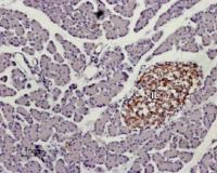
Supportive validation
- Submitted by
- OriGene (provider)
- Main image
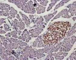
- Experimental details
- Expression of KISS1R in rat pancreas. Immunohistochemical staining of paraffin embedded section of rat pancreas using Anti-Kisspeptin Receptor (extracellular) antibody , (1:100). KISS1R staining (brown) appears in Isles of Langerhans (IL). Hematoxilin is used as the counterstain.
- Validation comment
- IHC
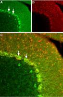
- Submitted by
- OriGene (provider)
- Main image

- Experimental details
- Expression of KISS1R in rat cerebellum Immunohistochemical staining of frozen rat cerebellum section using Anti-Kisspeptin Receptor (extracellular) antibody, (1:100). A. KISS1R (green) was expressed particularly in Purkinje cell bodies (arrows). B.Staining with mouse anti parvalbumin (red) detected Purkinje cells and interneurons in the molecular layer. C. Merge of the two images demonstrates that the staining was restricted to Purkinje cells.
- Validation comment
- IHC