Explore
 Validate
 Learn
Learn Western blot
Antibody data
- Antibody Data
- Antigen structure
- References [1]
- Comments [0]
- Validations
- Western blot [5]
- Immunocytochemistry [1]
- Immunohistochemistry [2]
- Flow cytometry [1]
Submit
Validation data
Reference
Comment
Report error
- Product number
- MA5-17096 - Provider product page

- Provider
- Invitrogen Antibodies
- Product name
- IGF2 Monoclonal Antibody (8H1)
- Antibody type
- Monoclonal
- Antigen
- Purifed from natural sources
- Description
- MA5-17096 targets IGF2 in FACS, ICC, IHC, IF and WB applications and shows reactivity with Human samples.
- Antibody clone number
- 8H1
- Concentration
- 1 mg/mL
Submitted references Non-islet Cell Hypoglycemia: Case Series and Review of the Literature.
Garla V, Sonani H, Palabindala V, Gomez-Sanchez C, Subauste J, Lien LF
Frontiers in endocrinology 2019;10:316
Frontiers in endocrinology 2019;10:316
No comments: Submit comment
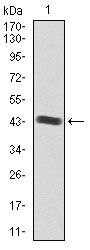
Supportive validation
- Submitted by
- Invitrogen Antibodies (provider)
- Main image

- Experimental details
- Western blot analysis of IGF2 using a IGF2 monoclonal antibody (Product # MA5-17096) against a human IGF2 recombinant protein.
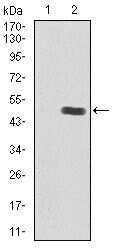
- Submitted by
- Invitrogen Antibodies (provider)
- Main image

- Experimental details
- Western blot analysis of IGF2 using IGF2 monoclonal antibody (Product # MA5-17096) in HEK293 (1) and IGF2 (AA: 25-180) human IgG Fc transfected HEK293 (2) cell lysate.
- Submitted by
- Invitrogen Antibodies (provider)
- Main image

- Experimental details
- Western blot analysis of IGF2 using a IGF2 monoclonal antibody (Product # MA5-17096) against a human IGF2 recombinant protein.
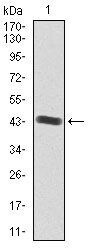
- Submitted by
- Invitrogen Antibodies (provider)
- Main image

- Experimental details
- Western blot analysis of IGF2 using IGF2 monoclonal antibody (Product # MA5-17096) in HEK293 (1) and IGF2 (AA: 25-180) human IgG Fc transfected HEK293 (2) cell lysate.

- Submitted by
- Invitrogen Antibodies (provider)
- Main image
- Experimental details
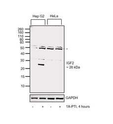
- Western blot was performed using Anti-IGF2 Monoclonal Antibody (8H1) (Product # MA5-17096) and a 26 kDa band corresponding to IGF2 was observed in Hep G2 (positive model) treated with PTI and not in PTI-treated HeLa (negative model). Whole cell extracts (30 µg lysate) of Hep G2 (Lane 1), Hep G2 treated with 1X-PTI for 4 hours (Lane 2), HeLa (Lane 3) and HeLa treated with 1X-PTI for 4 hours (Lane 4) were electrophoresed using NuPAGE™ 12% Bis-Tris Protein Gel (Product # NP0342BOX). Resolved proteins were then transferred onto a Nitrocellulose membrane (Product # IB23002) by iBlot® 2 Dry Blotting System (Product # IB21001). The blot was probed with the primary antibody (1:1000 dilution) and detected by chemiluminescence with Goat anti-Mouse IgG (H+L) Superclonal™ Recombinant Secondary Antibody, HRP (Product # A28177, 1:4000) using the iBright FL 1000 (Product # A32752). Chemiluminescent detection was performed using Novex® ECL Chemiluminescent Substrate Reagent Kit (Product # WP20005). Uncharacterized bands (*) were observed around 50-55 kDa.
Supportive validation
- Submitted by
- Invitrogen Antibodies (provider)
- Main image

- Experimental details
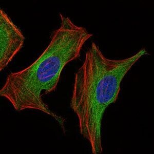
- Immunofluorescence analysis of HeLa cells using IGF2 monoclonal antibody (Product # MA5-17096) (Green). Blue: DRAQ5 fluorescent DNA dye. Red: actin filaments have been labeled with phalloidin.
Supportive validation
- Submitted by
- Invitrogen Antibodies (provider)
- Main image

- Experimental details
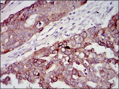
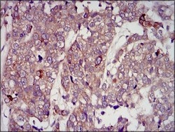
- Immunohistochemical analysis of paraffin-embedded ovarian cancer tissues using IGF2 monoclonal antibody (Product # MA5-17096) followed with DAB staining.
- Submitted by
- Invitrogen Antibodies (provider)
- Main image
- Experimental details
- Immunohistochemical analysis of paraffin-embedded bladder cancer tissues using IGF2 monoclonal antibody (Product # MA5-17096) followed with DAB staining.

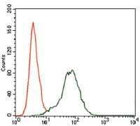
Supportive validation
- Submitted by
- Invitrogen Antibodies (provider)
- Main image

- Experimental details
- Flow cytometric analysis of HepG2 cells using IGF2 monoclonal antibody (Product # MA5-17096) (green) and negative control (red).