 Explore
 Validate
 Learn
Learn Western blot
Antibody data
- Antibody Data
- Antigen structure
- References [0]
- Comments [0]
- Validations
- Western blot [1]
- Immunohistochemistry [5]
Submit
Validation data
Reference
Comment
Report error
- Product number
- HPA042669 - Provider product page

- Provider
- Atlas Antibodies
- Proper citation
- Atlas Antibodies Cat#HPA042669, RRID:AB_10794916
- Product name
- Anti-TMEM259
- Antibody type
- Polyclonal
- Reactivity
- Human
- Host
- Rabbit
- Conjugate
- Unconjugated
- Antigen sequence
IKFELDIEPKVFKPPSSTEALNDSQEFPFPETPTK
VWPQDEYIVEYSLEYGFLRLSQATRQRLSIPVMVV
TL- Isotype
- IgG
- Vial size
- 100 µl
- Storage
- Store at +4°C for short term storage. Long time storage is recommended at -20°C.
No comments: Submit comment
Supportive validation
- Submitted by
- Atlas Antibodies (provider)
- Enhanced method
- Recombinant expression validation
- Main image

- Experimental details
- Western blot analysis in control (vector only transfected HEK293T lysate) and TMEM259 over-expression lysate (Co-expressed with a C-terminal myc-DDK tag (~3.1 kDa) in mammalian HEK293T cells, LY409545).

Supportive validation
- Submitted by
- Atlas Antibodies (provider)
- Main image
- Experimental details
- Immunohistochemical staining of human testis shows moderate cytoplasmic positivity in cells in seminiferous ducts and Leydig cells.
- Sample type
- HUMAN

- Submitted by
- Atlas Antibodies (provider)
- Main image
- Experimental details
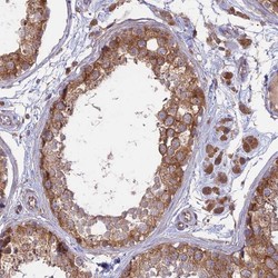
- Immunohistochemical staining of human prostate shows moderate granular cytoplasmic positivity in glandular cells.
- Sample type
- HUMAN

- Submitted by
- Atlas Antibodies (provider)
- Main image
- Experimental details
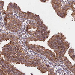
- Immunohistochemical staining of human Fallopian tube shows strong cytoplasmic positivity in glandular cells.
- Sample type
- HUMAN

- Submitted by
- Atlas Antibodies (provider)
- Main image

- Experimental details
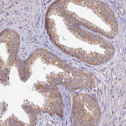
- Immunohistochemical staining of human testis shows strong granular cytoplasmic positivity in cells in seminiferous ducts.
- Sample type
- HUMAN

- Submitted by
- Atlas Antibodies (provider)
- Main image
- Experimental details
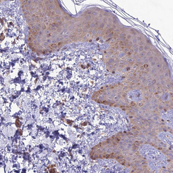
- Immunohistochemical staining of human skin shows weak cytoplasmic positivity in squamous epithelial cells.
- Sample type
- HUMAN
