Explore
 Validate
 Learn
Learn Immunocytochemistry
 Immunohistochemistry
Antibody data
- Antibody Data
- Antigen structure
- References [2]
- Comments [0]
- Validations
- Immunocytochemistry [1]
- Immunohistochemistry [4]
Submit
Validation data
Reference
Comment
Report error
- Product number
- HPA005464 - Provider product page

- Provider
- Atlas Antibodies
- Proper citation
- Atlas Antibodies Cat#HPA005464, RRID:AB_1848965
- Product name
- Anti-FMNL2
- Antibody type
- Polyclonal
- Reactivity
- Human
- Host
- Rabbit
- Conjugate
- Unconjugated
- Antigen sequence
ERVEELEENISHLSEKLQDTENEAMSKIVELEKQL
MQRNKELDVVREIYKDANTQVHTLRKMVKEKEEAI
QRQSTLEKKIHELEKQGTIKIQKKGDGDIAILPVV
ASGTLSMGSEVVAGNSVGP- Isotype
- IgG
- Vial size
- 100 µl
- Storage
- Store at +4°C for short term storage. Long time storage is recommended at -20°C.
Submitted references Junctional actin assembly is mediated by Formin-like 2 downstream of Rac1.
Characterization of Diaphanous-related formin FMNL2 in human tissues
Grikscheit K, Frank T, Wang Y, Grosse R
The Journal of cell biology 2015 May 11;209(3):367-76
The Journal of cell biology 2015 May 11;209(3):367-76
Characterization of Diaphanous-related formin FMNL2 in human tissues
Gardberg M, Talvinen K, Kaipio K, Iljin K, Kampf C, Uhlen M, Carpén O
BMC Cell Biology 2010 ;11(1):55
BMC Cell Biology 2010 ;11(1):55
No comments: Submit comment
Supportive validation
- Submitted by
- Atlas Antibodies (provider)
- Main image
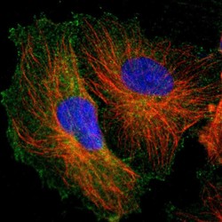
- Experimental details
- Immunofluorescent staining of human cell line U-251 MG shows localization to plasma membrane & cytosol.
- Sample type
- HUMAN
Enhanced validation
Supportive validation
- Submitted by
- Atlas Antibodies (provider)
- Enhanced method
- Orthogonal validation
- Main image
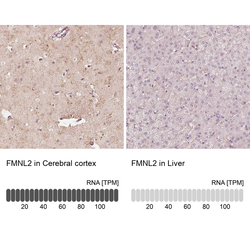
- Experimental details
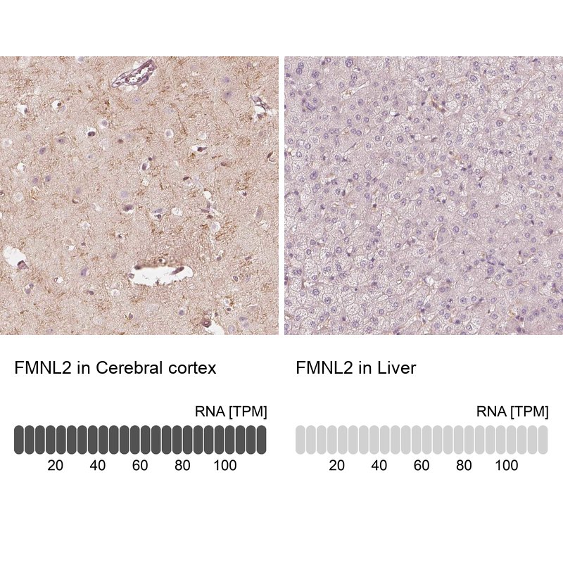
- Immunohistochemistry analysis in human cerebral cortex and liver tissues using Anti-FMNL2 antibody. Corresponding FMNL2 RNA-seq data are presented for the same tissues.
- Sample type
- HUMAN
Supportive validation
- Submitted by
- Atlas Antibodies (provider)
- Main image
- Experimental details
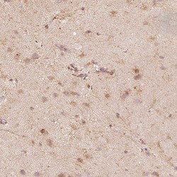
- Immunohistochemical staining of human lateral ventricle shows cytoplasmic positivity in neurons and glial cells.
- Submitted by
- Atlas Antibodies (provider)
- Main image
- Experimental details
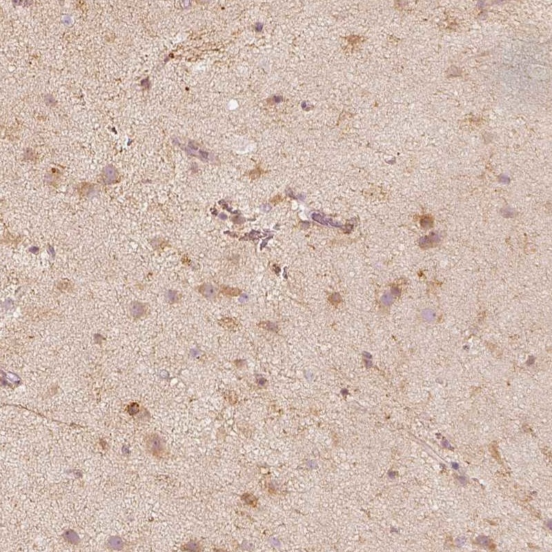
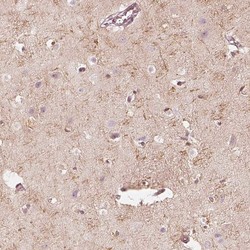
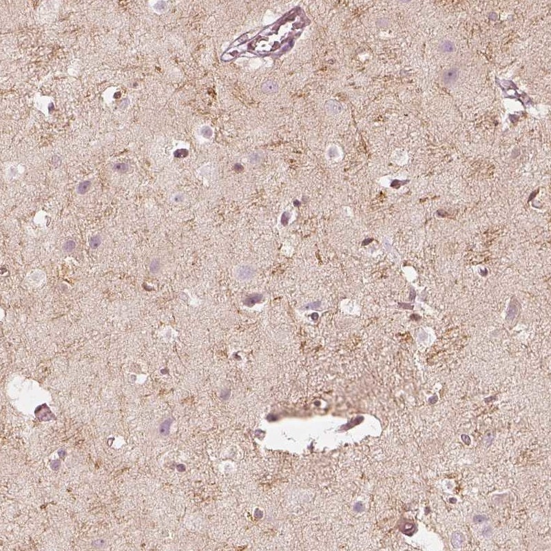
- Immunohistochemical staining of human cerebral cortex shows high expression.
- Sample type
- HUMAN
- Submitted by
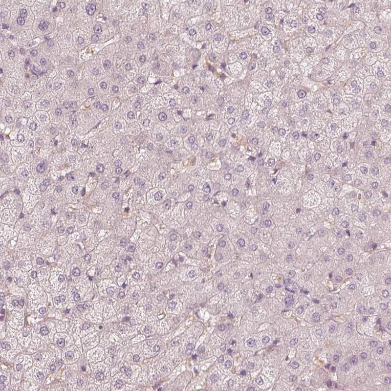
- Atlas Antibodies (provider)
- Main image
- Experimental details
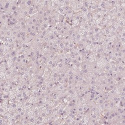
- Immunohistochemical staining of human liver shows low expression as expected.
- Sample type
- HUMAN