Explore
 Validate
 Learn
Learn Western blot
Antibody data
- Antibody Data
- Antigen structure
- References [3]
- Comments [0]
- Validations
- Western blot [3]
- Immunocytochemistry [1]
- Immunoprecipitation [1]
- Immunohistochemistry [2]
Submit
Validation data
Reference
Comment
Report error
- Product number
- GTX102340 - Provider product page

- Provider
- GeneTex
- Proper citation
- GeneTex Cat#GTX102340, RRID:AB_10618731
- Product name
- Calpain 1 antibody [N3C2], Internal
- Antibody type
- Polyclonal
- Reactivity
- Human, Mouse
- Host
- Rabbit
Submitted references Artocarpin induces cell apoptosis in human osteosarcoma cells through endoplasmic reticulum stress and reactive oxygen species.
The putative involvement of actin-binding proteins and cytoskeleton proteins in pathological mechanisms of ketamine cystitis-Revealed by a prospective pilot study using proteomic approaches.
Regulation of gene expression by NFAT transcription factors in hibernating ground squirrels is dependent on the cellular environment.
Lee CW, Chi MC, Chang TM, Liu JF
Journal of cellular physiology 2019 Aug;234(8):13157-13168
Journal of cellular physiology 2019 Aug;234(8):13157-13168
The putative involvement of actin-binding proteins and cytoskeleton proteins in pathological mechanisms of ketamine cystitis-Revealed by a prospective pilot study using proteomic approaches.
Yang HH, Zhai WJ, Kuo HC
Proteomics. Clinical applications 2017 Mar;11(3-4)
Proteomics. Clinical applications 2017 Mar;11(3-4)
Regulation of gene expression by NFAT transcription factors in hibernating ground squirrels is dependent on the cellular environment.
Zhang Y, Storey KB
Cell stress & chaperones 2016 Sep;21(5):883-94
Cell stress & chaperones 2016 Sep;21(5):883-94
No comments: Submit comment
Supportive validation
- Submitted by
- GeneTex (provider)
- Main image

- Experimental details
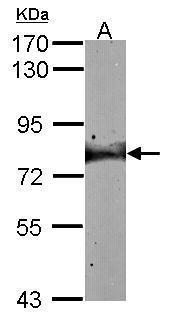
- Sample (30 ug of whole cell lysate) A: A431 (GTX27909) 7.5% SDS PAGE GTX102340 diluted at 1:1000
- Validation comment
- WB

- Submitted by
- GeneTex (provider)
- Main image

- Experimental details
- Sample (30 ug of whole cell lysate) A:NIH-3T3 7.5% SDS PAGE GTX102340 diluted at 1:1000
- Validation comment
- WB
- Submitted by
- GeneTex (provider)
- Main image
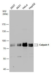
- Experimental details
- Calpain 1 antibody detects Calpain 1 protein by western blot analysis. Various whole cell extracts (30 ?g) were separated by 7.5% SDS-PAGE, and the membrane was blotted with Calpain 1 antibody (GTX102340) diluted at a dilution of 1:1000. The HRP-conjugated anti-rabbit IgG antibody (GTX213110-01) was used to detect the primary antibody.

Supportive validation
- Submitted by
- GeneTex (provider)
- Main image

- Experimental details
- Immunofluorescence analysis of methanol-fixed A431, using Calpain 1(GTX102340) antibody at 1:200 dilution.

Supportive validation
- Submitted by
- GeneTex (provider)
- Main image

- Experimental details
- Immunoprecipitation of Calpain 1 protein from A431 whole cell extracts using 5 £gg of Calpain 1 antibody [N3C2], Internal (GTX102340).Western blot analysis was performed using Calpain 1 antibody [N3C2], Internal (GTX102340).EasyBlot anti-Rabbit IgG (GTX221666-01) was used as a secondary reagent.

Supportive validation
- Submitted by
- GeneTex (provider)
- Main image
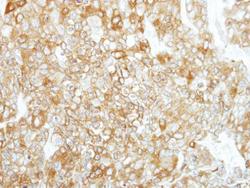
- Experimental details
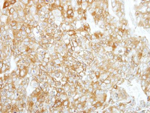
- Immunohistochemical analysis of paraffin-embedded BT474 xenograft, using Calpain 1(GTX102340) antibody at 1:500 dilution.
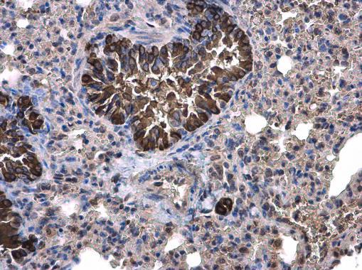
- Submitted by
- GeneTex (provider)
- Main image
- Experimental details
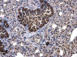
- Calpain 1 antibody [N3C2], Internal detects Calpain 1 protein at cytoplasm on mouse lung by immunohistochemical analysis. Sample: Paraffin-embedded mouse lung. Calpain 1 antibody [N3C2], Internal (GTX102340) diluted at 1:500.