Explore
 Validate
 Learn
LearnHPA002317
antibody from Atlas Antibodies
Targeting: BIRC3
API2, c-IAP2, cIAP2, hiap-1, MALT2, MIHC, RNF49
 Western blot
 Immunocytochemistry
Antibody data
- Antibody Data
- Antigen structure
- References [3]
- Comments [0]
- Validations
- Western blot [1]
- Immunocytochemistry [1]
- Immunohistochemistry [7]
Submit
Validation data
Reference
Comment
Report error
- Product number
- HPA002317 - Provider product page

- Provider
- Atlas Antibodies
- Proper citation
- Atlas Antibodies Cat#HPA002317, RRID:AB_1846748
- Product name
- Anti-BIRC3
- Antibody type
- Polyclonal
- Reactivity
- Human, Mouse, Rat
- Host
- Rabbit
- Conjugate
- Unconjugated
- Antigen sequence
QHAKWFPRCEYLIRIKGQEFIRQVQASYPHLLEQL
LSTSDSPGDENAESSIIHFEPGEDHSEDAIMMNTP
VINAAVEMGFSRSLVKQTVQRKILATGENYRLVND
LVLDLLNAEDEIREEERERATEE- Isotype
- IgG
- Vial size
- 100 µl
- Storage
- Store at +4°C for short term storage. Long time storage is recommended at -20°C.
Submitted references Phase I Trial of Induction Histone Deacetylase and Proteasome Inhibition Followed by Surgery in Non–Small-Cell Lung Cancer
Zoledronic acid directly suppresses cell proliferation and induces apoptosis in highly tumorigenic prostate and breast cancers.
Tissue profiling of the mammalian central nervous system using human antibody-based proteomics.
Jones D, Moskaluk C, Gillenwater H, Petroni G, Burks S, Philips J, Rehm P, Olazagasti J, Kozower B, Bao Y
Journal of Thoracic Oncology 2012 November;7(11):1683-1690
Journal of Thoracic Oncology 2012 November;7(11):1683-1690
Zoledronic acid directly suppresses cell proliferation and induces apoptosis in highly tumorigenic prostate and breast cancers.
Almubarak H, Jones A, Chaisuparat R, Zhang M, Meiller TF, Scheper MA
Journal of carcinogenesis 2011 Jan 15;10:2
Journal of carcinogenesis 2011 Jan 15;10:2
Tissue profiling of the mammalian central nervous system using human antibody-based proteomics.
Mulder J, Björling E, Jonasson K, Wernérus H, Hober S, Hökfelt T, Uhlén M
Molecular & cellular proteomics : MCP 2009 Jul;8(7):1612-22
Molecular & cellular proteomics : MCP 2009 Jul;8(7):1612-22
No comments: Submit comment
Enhanced validation
- Submitted by
- Atlas Antibodies (provider)
- Enhanced method
- Recombinant expression validation
- Main image

- Experimental details
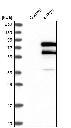
- Western blot analysis in control (vector only transfected HEK293T lysate) and BIRC3 over-expression lysate (Co-expressed with a C-terminal myc-DDK tag (~3.1 kDa) in mammalian HEK293T cells, LY420093).
Supportive validation
- Submitted by
- Atlas Antibodies (provider)
- Main image
- Experimental details
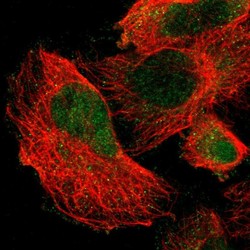
- Immunofluorescent staining of human cell line U-251 MG shows localization to nucleoplasm & cytosol.
- Sample type
- HUMAN

Supportive validation
- Submitted by
- Atlas Antibodies (provider)
- Main image

- Experimental details
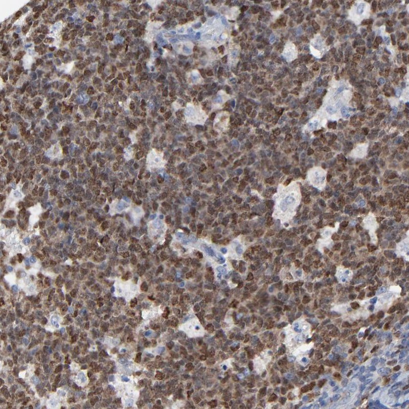
- Immunohistochemical staining of human tonsil shows nuclear positivity in reaction center cells.
- Submitted by
- Atlas Antibodies (provider)
- Main image
- Experimental details
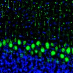
- Immunofluorescence staining of mouse cerebellum shows strong immunoreactivity in Purkinje cells.
- Sample type
- HUMAN
- Submitted by
- Atlas Antibodies (provider)
- Main image
- Experimental details
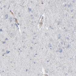
- Immunohistochemical staining of human cerebral cortex shows positivity in endothelial cells.

- Submitted by
- Atlas Antibodies (provider)
- Main image

- Experimental details
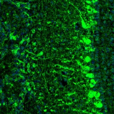
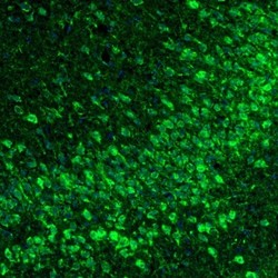
- Immunofluorescence staining of mouse olfactory bulb shows cytoplasmic staining in mitral and external plexiform cell layers.
- Sample type
- HUMAN
- Submitted by
- Atlas Antibodies (provider)
- Main image
- Experimental details
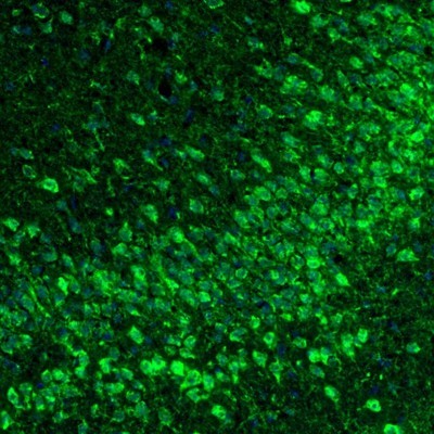
- Immunofluorescence staining of mouse piriform cortex shows cytoplasmic positivity in neurons.
- Sample type
- HUMAN
- Submitted by
- Atlas Antibodies (provider)
- Main image
- Experimental details
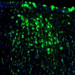
- Immunofluorescence staining of mouse thalamus shows neuronal staining in paraventricular nucleus.
- Sample type
- HUMAN

- Submitted by
- Atlas Antibodies (provider)
- Main image
- Experimental details
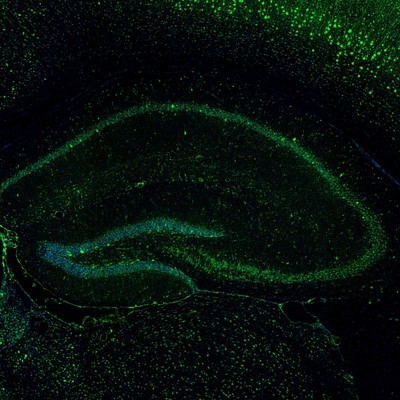
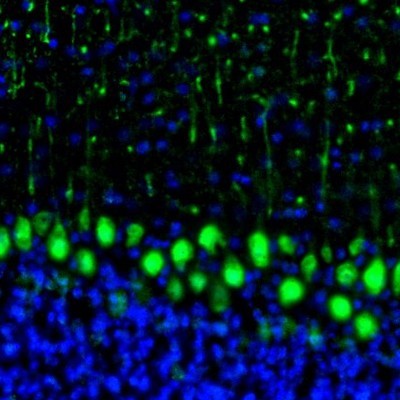
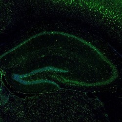
- Immunofluorescence staining of mouse hippocampus shows cytoplasmic positivity in a subset of neurons.
- Sample type
- HUMAN