Explore
 Validate
 Learn
LearnPA5-95497
antibody from Invitrogen Antibodies
Targeting: BIRC2
API1, c-IAP1, cIAP1, hiap-2, MIHB, RNF48
 Western blot
 ELISA
ELISAAntibody data
- Antibody Data
- Antigen structure
- References [0]
- Comments [0]
- Validations
- Western blot [1]
- Immunocytochemistry [1]
- Flow cytometry [2]
Submit
Validation data
Reference
Comment
Report error
- Product number
- PA5-95497 - Provider product page

- Provider
- Invitrogen Antibodies
- Product name
- cIAP1 Polyclonal Antibody
- Antibody type
- Polyclonal
- Antigen
- Recombinant full-length protein
- Reactivity
- Human, Mouse, Rat
- Host
- Rabbit
- Isotype
- IgG
- Vial size
- 100 µg
- Concentration
- 500 µg/mL
- Storage
- Store at 4°C short term. For long term storage, store at -20°C, avoiding freeze/thaw cycles.
No comments: Submit comment
Supportive validation
- Submitted by
- Invitrogen Antibodies (provider)
- Main image

- Experimental details
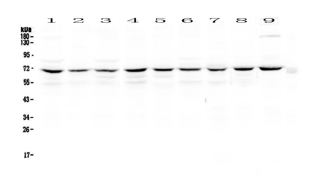
- Western blot analysis of cIAP1 in Lane 1: human HeLa whole cell lysates, Lane 2: human Jurkat whole cell lysates, Lane 3: human MCF-7 whole cell lysates, Lane 4: human COLO-320 whole cell lysates, Lane 5: human U-87MG whole cell lysates, Lane 6: human A549 whole cell lysates, Lane 7: rat thymus tissue lysates, Lane 8: mouse thymus tissue lysates, Lane 9: mouse testis tissue lysates. Electrophoresis was performed with 5-20% SDS-PAGE gel (70V, Stacking gel; 90V Resolving gel, Time: 2-3 hours), transferred to a nitrocellulose membrane and blocked using 5% Non-fat Milk/TBS (1.5 hrs at room temperature). Samples were incubated with cIAP1 polyclonal antibody (Product # PA5-95497) using a 0.5 µg/mL dilution, followed by a goat anti-rabbit IgG-HRP at a dilution of 1:10,000, and developed with enhanced chemiluminescence (ECL).
Supportive validation
- Submitted by
- Invitrogen Antibodies (provider)
- Main image
- Experimental details
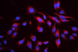
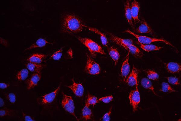
- Immunocytochemistry analysis of cIAP1 using anti-cIAP1 antibody (Product # PA5-95497). cIAP1 was detected in a section of U2OS cells. Enzyme antigen retrieval was performed using IHC enzyme antigen retrieval reagent for 15 mins. The cells were blocked with 10% goat serum and then incubated with 2μg/mL rabbit anti-cIAP1 antibody (Product # PA5-95497) overnight at 4°C. Cy3 Conjugated Goat Anti-Rabbit IgG was used as secondary antibody at 1:100 dilution and incubated for 30 minutes at 37°C. The section was counterstained with DAPI. Visualize using a fluorescence microscope and filter sets appropriate for the label used.
Supportive validation
- Submitted by
- Invitrogen Antibodies (provider)
- Main image
- Experimental details
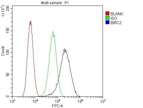
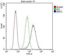
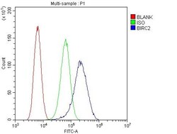
- Flow cytometry of cIAP1 in U20S cells (blue line), isotype control rabbit IgG (green line) and unlabeled (red line). Samples were blocked with 10% goat serum, incubated with cIAP1 polyclonal antibody (Product # PA5-95497) at a dilution of 1 µg (per 1x10^6 cells), followed by 488 conjugated goat anti-rabbit IgG (30 min at 20°C) using a 5-10 µg (per 1x10^6 cells) dilution.
- Submitted by
- Invitrogen Antibodies (provider)
- Main image
- Experimental details
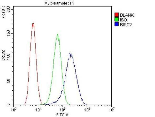
- Flow Cytometry of cIAP1 in U2OS cells (blue line), isotype control rabbit IgG (green line) and unlabeled (red line). Samples were blocked with 10% goat serum, incubated with cIAP1 Polyclonal Antibody (Product # PA5-95497) at a dilution of 1 μg (per 1x10^6 cells), followed by DyLight®488 conjugated goat anti-rabbit IgG (for 30 minutes at 20°C) using 5-10 μg (per 1x10^6 cells) dilution.