 Explore
 Validate
 Learn
Learn Western blot
Antibody data
- Antibody Data
- Antigen structure
- References [1]
- Comments [0]
- Validations
- Western blot [3]
Submit
Validation data
Reference
Comment
Report error
- Product number
- MAB818 - Provider product page

- Provider
- R&D Systems
- Product name
- Human cIAP-1/HIAP-2 Antibody
- Antibody type
- Monoclonal
- Description
- Protein A or G purified from hybridoma culture supernatant. Detects human cIAP-1/HIAP-2 in direct ELISAs and Western blots. In direct ELISAs, 100% cross-reactivity with recombinant human (rh) cIAP-2 (aa 2-604) and no cross-reactivity with rhBIRC6 (aa 4582-4735), rhcIAP-2 (aa 94-178), rhXIAP (aa 1-497), rhXIAP (BIR2 domain; aa 124-242), or rhXIAP (BIR3 domain; aa 252-356) is observed.
- Reactivity
- Human
- Host
- Mouse
- Conjugate
- Unconjugated
- Antigen sequence
Q13490- Isotype
- IgG
- Antibody clone number
- 681732
- Vial size
- 100 ug
- Concentration
- LYOPH
- Storage
- Use a manual defrost freezer and avoid repeated freeze-thaw cycles. 12 months from date of receipt, -20 to -70 °C as supplied. 1 month, 2 to 8 °C under sterile conditions after reconstitution. 6 months, -20 to -70 °C under sterile conditions after reconstitution.
Submitted references A novel function of cIAP1 as a mediator of CHIP-driven eIF4E regulation.
Seo TW, Lee JS, Choi YN, Jeong DH, Lee SK, Yoo SJ
Scientific reports 2017 Aug 29;7(1):9816
Scientific reports 2017 Aug 29;7(1):9816
No comments: Submit comment
Supportive validation
- Submitted by
- R&D Systems (provider)
- Main image
- Experimental details
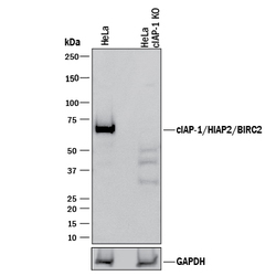
- Western Blot Shows Human cIAP-1/HIAP-2 Specificity by Using Knockout Cell Line. Western blot shows lysates of HeLa human cervical epithelial carcinoma parental cell line and cIAP-1/HIAP-2 knockout HeLa cell line (KO). PVDF membrane was probed with 1 µg/mL of Mouse Anti-Human cIAP-1/HIAP-2 Monoclonal Antibody (Catalog # MAB818) followed by HRP-conjugated Anti-Mouse IgG Secondary Antibody (Catalog # HAF018). A specific band was detected for cIAP-1/HIAP-2 at approximately 68 kDa (as indicated) in the parental HeLa cell line, but is not detectable in knockout HeLa cell line. GAPDH (Catalog # MAB5718) is shown as a loading control. This experiment was conducted under reducing conditions and using Immunoblot Buffer Group 1.

- Submitted by
- R&D Systems (provider)
- Main image
- Experimental details
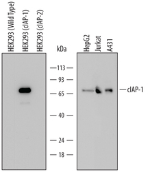
- Detection of Human cIAP-1/HIAP-2 by Western Blot. Western blot shows lysates of HEK293 human embryonic kidney cell line either mock transfected, transfected with full length human cIAP-1, or transfected with full length human cIAP-2, HepG2 human hepatocellular carcinoma cell line, Jurkat human acute T cell leukemia cell line, and A431 human epithelial carcinoma cell line. PVDF Membrane was probed with 1 µg/mL of Human cIAP-1/HIAP-2 Monoclonal Antibody (Catalog # MAB818) followed by HRP-conjugated Anti-Mouse IgG Secondary Antibody (Catalog # HAF007). A specific band was detected for cIAP-1/HIAP-2 at approximately 72 kDa (as indicated). This experiment was conducted under reducing conditions and using Immunoblot Buffer Group 2.

- Submitted by
- R&D Systems (provider)
- Main image
- Experimental details
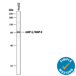
- Detection of Human cIAP-1/HIAP-2 by Simple WesternTM. Simple Western lane view shows lysates of HepG2 human hepatocellular carcinoma cell line, loaded at 0.2 mg/mL. A specific band was detected for cIAP-1/HIAP-2 at approximately 66 kDa (as indicated) using 5 µg/mL of Mouse Anti-Human cIAP-1/HIAP-2 Monoclonal Antibody (Catalog # MAB818) . This experiment was conducted under reducing conditions and using the 12-230 kDa separation system.
