 Explore
 Validate
 Learn
Learn Western blot
 ELISA
ELISAAntibody data
- Antibody Data
- Antigen structure
- References [2]
- Comments [0]
- Validations
- Western blot [3]
Submit
Validation data
Reference
Comment
Report error
- Product number
- NBP2-29420 - Provider product page

- Provider
- Novus Biologicals
- Product name
- Mouse Monoclonal UCH-L1/PGP9.5 Antibody
- Antibody type
- Monoclonal
- Description
- Protein G purified. This MAb reacts with a protein of 20-30kDa, identified as PGP9.5, also known as ubiquitin carboxyl-terminal hydrolase-1 (UchL1). Initially, PGP9.5 expression in normal tissues was reported in neurons and neuroendocrine cells but later it was found in distal renal tubular epithelium, spermatogonia, Leydig cells, oocytes, melanocytes, prostatic secretory epithelium, ejaculatory duct cells, epididymis, mammary epithelial cells, Merkel cells, and dermal fibroblasts. Furthermore, immunostaining for PGP9.5 has been shown in a wide variety of mesenchymal neoplasms as well. A mutation in PGP9.5 gene is believed to cause a form of Parkinson's disease.
- Reactivity
- Human, Mouse, Rat, Bovine, Canine, Guinea Pig, Porcine, Rabbit, Sheep, Zebrafish
- Host
- Mouse
- Isotype
- IgG
- Vial size
- 0.1 mg
- Concentration
- 0.2 mg/ml
- Storage
- Store at 4C.
Submitted references Suitability of antigens PGP 9.5 and neurofilament light as marker proteins for detection of neuronal tissue in processed meat products.
Suitability of antigens PGP 9.5 and neurofilament light as marker proteins for detection of neuronal tissue in processed meat products.
Gaunitz C, Gabert J, Lücker E, Seeger J, Stahl T
Journal of food protection 2009 May;72(5):1070-7
Journal of food protection 2009 May;72(5):1070-7
Suitability of antigens PGP 9.5 and neurofilament light as marker proteins for detection of neuronal tissue in processed meat products.
Gaunitz C, Gabert J, Lücker E, Seeger J, Stahl T
Journal of food protection 2009 May;72(5):1070-7
Journal of food protection 2009 May;72(5):1070-7
No comments: Submit comment
Supportive validation
- Submitted by
- Novus Biologicals (provider)
- Main image

- Experimental details
- Simple Western: UCH-L1/PGP9.5 Antibody (13C4) [NBP2-29420] - Simple Western lane view shows a specific band for PGP9.5 / UCHL-1 in 0.2 mg/ml of h. Cerebellum (left) and IMR-32 (right) lysate(s). This experiment was performed under reducing conditions using the 12-230 kDa separation system.

- Submitted by
- Novus Biologicals (provider)
- Main image
- Experimental details
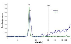
- Simple Western: UCH-L1/PGP9.5 Antibody (13C4) [NBP2-29420] - Electropherogram image of the corresponding Simple Western lane. PGP9.5 / UCHL-1 antibody was used at 10 ug/ml dilution of h. Cerebellum and IMR-32 lysates(s) respectively.

- Submitted by
- Novus Biologicals (provider)
- Main image

- Experimental details
- Western Blot: UCH-L1/PGP9.5 Antibody (13C4) [NBP2-29420] - Western Blot Analysis of human brain tissue lysate using UCH-L1/PGP9.5 Antibody (13C4)
