 Explore
 Validate
 Learn
Learn Western blot
Antibody data
- Antibody Data
- Antigen structure
- References [4]
- Comments [0]
- Validations
- Western blot [1]
- Immunohistochemistry [1]
Submit
Validation data
Reference
Comment
Report error
- Product number
- MAB1100 - Provider product page

- Provider
- Abnova Corporation
- Proper citation
- Abnova Corporation Cat#MAB1100, RRID:AB_1581669
- Product name
- UCHL1 monoclonal antibody, clone 3D9
- Antibody type
- Monoclonal
- Description
- Mouse monoclonal antibody raised against full length recombinant UCHL1.
- Isotype
- IgG
- Antibody clone number
- 3D9
- Storage
- Store at -20°C.Aliquot to avoid repeated freezing and thawing.
Submitted references Immunohistochemical analysis of neuropeptides (protein gene product 9.5, substance P and calcitonin gene-related peptide) in hypertrophic burn scar with pain and itching.
Ubiquitin C-terminal hydrolase L1 regulates the morphology of neural progenitor cells and modulates their differentiation.
The UCH-L1 gene encodes two opposing enzymatic activities that affect alpha-synuclein degradation and Parkinson's disease susceptibility.
PGP9.5 as a candidate tumor marker for non-small-cell lung cancer.
Kwak IS, Choi YH, Jang YC, Lee YK
Burns : journal of the International Society for Burn Injuries 2014 Dec;40(8):1661-7
Burns : journal of the International Society for Burn Injuries 2014 Dec;40(8):1661-7
Ubiquitin C-terminal hydrolase L1 regulates the morphology of neural progenitor cells and modulates their differentiation.
Sakurai M, Ayukawa K, Setsuie R, Nishikawa K, Hara Y, Ohashi H, Nishimoto M, Abe T, Kudo Y, Sekiguchi M, Sato Y, Aoki S, Noda M, Wada K
Journal of cell science 2006 Jan 1;119(Pt 1):162-71
Journal of cell science 2006 Jan 1;119(Pt 1):162-71
The UCH-L1 gene encodes two opposing enzymatic activities that affect alpha-synuclein degradation and Parkinson's disease susceptibility.
Liu Y, Fallon L, Lashuel HA, Liu Z, Lansbury PT Jr
Cell 2002 Oct 18;111(2):209-18
Cell 2002 Oct 18;111(2):209-18
PGP9.5 as a candidate tumor marker for non-small-cell lung cancer.
Hibi K, Westra WH, Borges M, Goodman S, Sidransky D, Jen J
The American journal of pathology 1999 Sep;155(3):711-5
The American journal of pathology 1999 Sep;155(3):711-5
No comments: Submit comment
Supportive validation
- Submitted by
- Abnova Corporation (provider)
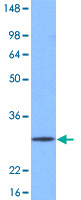
- Main image

- Experimental details
- Western blot analysis of mouse brain extracts (20 ug) were resolved by SDS - PAGE , transferred to NC membrane and probed with UCHL1 monoclonal antibody , clone 3D9 (1 : 1000) (Cat # MAB1100). Proteins were visualized using a goat anti - mouse secondary antibody conjugated to HRP and an ECL detection system.
Supportive validation
- Submitted by
- Abnova Corporation (provider)
- Main image
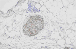
- Experimental details
- Paraffin embedded sections of human nerve tissue were incubated with UCHL1 monoclonal antibody , clone 3D9 (Cat # MAB1100) (1:100) for 2 hours at room temperature. Antigen retrieval was performed in 0.1M sodium citrate buffer and detected using Diaminobenzidine (DAB).
- Validation comment
- Immunohistochemistry (Formalin/PFA-fixed paraffin-embedded sections)
