Antibody data
- Antibody Data
- Antigen structure
- References [11]
- Comments [0]
- Validations
- Western blot [3]
- Immunocytochemistry [3]
- Immunohistochemistry [2]
- Other assay [17]
Submit
Validation data
Reference
Comment
Report error
- Product number
- 480012 - Provider product page

- Provider
- Invitrogen Antibodies
- Product name
- PGP9.5 Monoclonal Antibody (BH7)
- Antibody type
- Monoclonal
- Antigen
- Purifed from natural sources
- Reactivity
- Human, Mouse, Rat, Bovine, Porcine
- Host
- Mouse
- Isotype
- IgG
- Antibody clone number
- BH7
- Vial size
- 100 µL
- Concentration
- Conc. Not Determined
- Storage
- -20° C, Avoid Freeze/Thaw Cycles
Submitted references Effects of Different n6/n3 PUFAs Dietary Ratio on Cardiac Diabetic Neuropathy.
Expression of Connexins 37, 43 and 45 in Developing Human Spinal Cord and Ganglia.
Reactive-site-centric chemoproteomics identifies a distinct class of deubiquitinase enzymes.
MicroRNA-922 promotes tau phosphorylation by downregulating ubiquitin carboxy-terminal hydrolase L1 (UCHL1) expression in the pathogenesis of Alzheimer's disease.
Cutaneous expression of calcium/calmodulin-dependent protein kinase II in rats with type 1 and type 2 diabetes.
Gender and gonadectomy influence on neurons in superior cervical ganglia of sexually mature rats.
Changes in epidermal thickness and cutaneous innervation during maturation in long-term diabetes.
Reduced epidermal thickness, nerve degeneration and increased pain-related behavior in rats with diabetes type 1 and 2.
Conformational stabilization of ubiquitin yields potent and selective inhibitors of USP7.
Immunofluorescent triple-staining technique to identify sensory nerve endings in human thumb ligaments.
Ubiquitin editing enzyme UCH L1 and microtubule dynamics: implication in mitosis.
Urlić M, Urlić I, Urlić H, Mašek T, Benzon B, Vitlov Uljević M, Vukojević K, Filipović N
Nutrients 2020 Sep 10;12(9)
Nutrients 2020 Sep 10;12(9)
Expression of Connexins 37, 43 and 45 in Developing Human Spinal Cord and Ganglia.
Jurić M, Zeitler J, Vukojević K, Bočina I, Grobe M, Kretzschmar G, Saraga-Babić M, Filipović N
International journal of molecular sciences 2020 Dec 8;21(24)
International journal of molecular sciences 2020 Dec 8;21(24)
Reactive-site-centric chemoproteomics identifies a distinct class of deubiquitinase enzymes.
Hewings DS, Heideker J, Ma TP, AhYoung AP, El Oualid F, Amore A, Costakes GT, Kirchhofer D, Brasher B, Pillow T, Popovych N, Maurer T, Schwerdtfeger C, Forrest WF, Yu K, Flygare J, Bogyo M, Wertz IE
Nature communications 2018 Mar 21;9(1):1162
Nature communications 2018 Mar 21;9(1):1162
MicroRNA-922 promotes tau phosphorylation by downregulating ubiquitin carboxy-terminal hydrolase L1 (UCHL1) expression in the pathogenesis of Alzheimer's disease.
Zhao ZB, Wu L, Xiong R, Wang LL, Zhang B, Wang C, Li H, Liang L, Chen SD
Neuroscience 2014 Sep 5;275:232-7
Neuroscience 2014 Sep 5;275:232-7
Cutaneous expression of calcium/calmodulin-dependent protein kinase II in rats with type 1 and type 2 diabetes.
Boric M, Jelicic Kadic A, Puljak L
Journal of chemical neuroanatomy 2014 Nov;61-62:140-6
Journal of chemical neuroanatomy 2014 Nov;61-62:140-6
Gender and gonadectomy influence on neurons in superior cervical ganglia of sexually mature rats.
Filipović N, Zuvan L, Mašek T, Tokalić R, Grković I
Neuroscience letters 2014 Mar 20;563:55-60
Neuroscience letters 2014 Mar 20;563:55-60
Changes in epidermal thickness and cutaneous innervation during maturation in long-term diabetes.
Jelicic Kadic A, Boric M, Vidak M, Ferhatovic L, Puljak L
Journal of tissue viability 2014 Feb;23(1):7-12
Journal of tissue viability 2014 Feb;23(1):7-12
Reduced epidermal thickness, nerve degeneration and increased pain-related behavior in rats with diabetes type 1 and 2.
Boric M, Skopljanac I, Ferhatovic L, Jelicic Kadic A, Banozic A, Puljak L
Journal of chemical neuroanatomy 2013 Nov;53:33-40
Journal of chemical neuroanatomy 2013 Nov;53:33-40
Conformational stabilization of ubiquitin yields potent and selective inhibitors of USP7.
Zhang Y, Zhou L, Rouge L, Phillips AH, Lam C, Liu P, Sandoval W, Helgason E, Murray JM, Wertz IE, Corn JE
Nature chemical biology 2013 Jan;9(1):51-8
Nature chemical biology 2013 Jan;9(1):51-8
Immunofluorescent triple-staining technique to identify sensory nerve endings in human thumb ligaments.
Lee J, Ladd A, Hagert E
Cells, tissues, organs 2012;195(5):456-64
Cells, tissues, organs 2012;195(5):456-64
Ubiquitin editing enzyme UCH L1 and microtubule dynamics: implication in mitosis.
Bheda A, Gullapalli A, Caplow M, Pagano JS, Shackelford J
Cell cycle (Georgetown, Tex.) 2010 Mar 1;9(5):980-94
Cell cycle (Georgetown, Tex.) 2010 Mar 1;9(5):980-94
No comments: Submit comment
Supportive validation
- Submitted by
- Invitrogen Antibodies (provider)
- Main image

- Experimental details
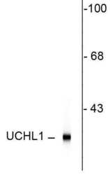
- Western blot of rat hippocampal homogenate showing specific immunolabeling of the ~ 24k UCHL1 protein.
- Submitted by
- Invitrogen Antibodies (provider)
- Main image

- Experimental details
- Western blot was performed using Anti-PGP9.5 Monoclonal Antibody (BH7) (Product # 480012) and a 24 kDa band corresponding to PGP9.5 was observed in DU 145, SH-SY5Y, Neuro-2A, RSC96, Rat Brain and Mouse Brain. Whole cell extracts (30 µg lysate) of DU 145 (Lane 1), SH-SY5Y (Lane 2), K-562 (Lane 3), HeLa (Lane 4), Hep G2 (Lane 5), Neuro-2A (Lane 6), RSC96 (Lane 7) and tissue extracts of Rat Brain (Lane 8), Rat Liver (Lane 9), Rat Heart (Lane 10) and Mouse Brain (Lane 11) were electrophoresed using NuPAGE™ 12% Bis-Tris Protein Gel (Product # NP0342BOX). Resolved proteins were then transferred onto a nitrocellulose membrane (Product # IB23001) by iBlot® 2 Dry Blotting System (Product # IB21001). The blot was probed with the primary antibody (1:5000 dilution) and detected by chemiluminescence with Goat anti-Mouse IgG (H+L), Superclonal™ Recombinant Secondary Antibody, HRP (Product # A28177, 1:4000 dilution) using the iBright FL 1000 (Product # A32752). Chemiluminescent detection was performed using Novex® ECL Chemiluminescent Substrate Reagent Kit (Product # WP20005).
- Submitted by
- Invitrogen Antibodies (provider)
- Main image

- Experimental details
- Western blot of PGP9.5 in rat hippocampal homogenate showing specific immunolabeling of a ~24 kDa band corresponding to PGP9.5 monoclonal antibody (Product # 480012).
Supportive validation
- Submitted by
- Invitrogen Antibodies (provider)
- Main image

- Experimental details
- Rat spinal cord stained with anti-UCHL1 (red) and anti-neurofilament NF-H antibody (green). The large cells are α-motorneurons and UCHL1 fills the cytoplasm of their perikarya and dendrites.
- Submitted by
- Invitrogen Antibodies (provider)
- Main image

- Experimental details
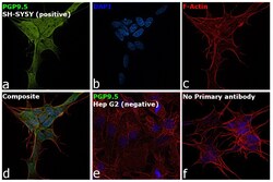
- Immunofluorescence analysis of PGP9.5 was performed using 70% confluent log phase SH-SY5Y cells. The cells were fixed with 4% paraformaldehyde for 10 minutes, permeabilized with 0.1% Triton™ X-100 for 10 minutes, and blocked with 2% BSA for 45 minutes at room temperature. The cells were labeled with PGP9.5 Monoclonal Antibody (BH7) (Product # 480012) at 1:500 dilution in 0.1% BSA, incubated at 4 degree celsius overnight and then labeled with Donkey anti-Mouse IgG (H+L) Highly Cross-Adsorbed Secondary Antibody, Alexa Fluor Plus 488 (Product # A32766, 1:2000 dilution) for 45 minutes at room temperature (Panel a: Green). Nuclei (Panel b: Blue) were stained with ProLong™ Diamond Antifade Mountant with DAPI (Product # P36962). F-actin (Panel c: Red) was stained with Rhodamine Phalloidin (Product # R415, 1:300 dilution). Panel d represents the merged image showing nuclear and cytoplasmic localization. Panel e represents Hep G2 cells having no expression of PGP9.5 Panel f represents control cells with no primary antibody to assess background. The images were captured at 60X magnification.
- Submitted by
- Invitrogen Antibodies (provider)
- Main image

- Experimental details
- Immunocytochemistry analysis of PGP9.5 in HEK 293 cells labeled with PGP9.5 monoclonal antibody (Product # 480012) using a dilution of 1:500 (green) and Anti-Neuron specific enolase antibody using a dilution of 1:500 (red). The blue is staining nuclear DNA.
Supportive validation
- Submitted by
- Invitrogen Antibodies (provider)
- Main image

- Experimental details
- Immunohistochemistry analysis of PGP9.5 in rat hippocampus showing specific labeling of PGP9.5 monoclonal antibody (Product # 480012) using a dilution of 1:5,000 (green) in cell bodies, dendrites of neurons, and specific labeling of FOX3(red). The blue is DAPI staining of nuclear DNA.
- Submitted by
- Invitrogen Antibodies (provider)
- Main image

- Experimental details
- Immunohistochemistry analysis of PGP9.5 in rat spinal cord labeled with PGP9.5 monoclonal antibody (Product # 480012) using a dilution of 1:500 (red) and anti-Neurofilament H with a dilution of 1:25,000 (green). The large cells are alpha-Motorneurons and UCHL1 fills the cytoplasm of their perikarya and dendrites.
Supportive validation
- Submitted by
- Invitrogen Antibodies (provider)
- Main image

- Experimental details
- NULL
- Submitted by
- Invitrogen Antibodies (provider)
- Main image

- Experimental details
- NULL
- Submitted by
- Invitrogen Antibodies (provider)
- Main image

- Experimental details
- NULL
- Submitted by
- Invitrogen Antibodies (provider)
- Main image

- Experimental details
- NULL
- Submitted by
- Invitrogen Antibodies (provider)
- Main image

- Experimental details
- NULL
- Submitted by
- Invitrogen Antibodies (provider)
- Main image

- Experimental details
- NULL
- Submitted by
- Invitrogen Antibodies (provider)
- Main image

- Experimental details
- NULL
- Submitted by
- Invitrogen Antibodies (provider)
- Main image

- Experimental details
- NULL
- Submitted by
- Invitrogen Antibodies (provider)
- Main image

- Experimental details
- NULL
- Submitted by
- Invitrogen Antibodies (provider)
- Main image

- Experimental details
- NULL
- Submitted by
- Invitrogen Antibodies (provider)
- Main image

- Experimental details
- Figure 1 Cardiac weight, body weight changes and sampling of visual fields. ( A ) Cardiac weight (CW; g) of rats in different experimental groups: c--control group; stz--diabetic group fed standard diet (both diets n6/n3 ratio 7); stz+DHA--diabetic group supplemented with n3 polyunsaturated fatty acid (PUFA) (2.5% of fish oil, containing 16% eicosapentaenoic acid--EPA and 19% docosahexaenoic acid--DHA; n6/n3 ratio of 1); stz+n6--Diabetic group fed with diet that contained 2.5% sunflower oil (n6/n3 ratio 60). *-- p < 0.05; **-- p < 0.01 difference between indicated groups. ( B ) Cardiac weight expressed as percentage of body weight (%BW). No significant difference in CW expressed as %BW was found (ns). ( C ) Body weight increase or decrease (BW I/D) expressed as % of the initial weight. ( D ) Correlation analysis between CW (g), CW (%BW) and BW I/D (%)--Pearson's coefficient of correlation (r). ( E ) section through the ventricles of the rat heart (non-related specimen), showing example of positioning of the visual fields for analysis--M--Intramural (Mid.) part of the cardiac septum; R--Right subendocardial area of the septum; L--Left subendocardial area of the septum. ( F ) Representative photomicrographs showing comparison of the density of fibers in the right side of the subendocardial area of the cardiac septum (F1 and F2) and Mid. area (F3 and F4) of the same animal, stained for general neuronal marker Protein g Product (PgP) 9.5 (green). Tresholded figures F2 (tresholded
- Submitted by
- Invitrogen Antibodies (provider)
- Main image

- Experimental details
- Figure 2 Protein g Product 9.5 immunoreactive nerve fiber density in subendocardial areas of cardiac septum. ( A ) Representative photomicrographs of right side of the subendocardial area of the cardiac septum stained for general neuronal marker PgP 9.5 (green). c--control group; stz--diabetic group fed standard diet (both diets n6/n3 ratio 7); stz+DHA--diabetic group supplemented with n3 PUFA (2.5% of fish oil, containing 16% eicosapentaenoic acid--EPA and 19% docosahexaenoic acid--DHA; n6/n3 ratio of 1); stz+n6--Diabetic group fed with diet that contained 2.5% sunflower oil (n6/n3 ratio 60). ( B ) Threshold figures from A; white--nerve fiber area. ( C ) Neuronal density expressed as number of fibers per area unit. L--Subendocardial area facing the left ventricle; R--Subendocardial area facing right ventricle; L+R--Average from L and R. ( D ) --Neuronal density expressed as percentage of the tissue area. *-- p < 0.05 difference between indicated groups. Scale-bar = 20 um.
- Submitted by
- Invitrogen Antibodies (provider)
- Main image

- Experimental details
- Figure 3 Expression of Cxs 37, 43, and 45 in the inner layer (INL) of the SC in human conceptuses. Thoracic segments of the SC of 6, 7.5, and 10 week-old human conceptuses were stained for Cx37, Cx43, and Cx45 (green), and protein gene peptide (PGP) 9.5 (red). The INL was presented on photomicrographs, with neuroepithelium (ne)--a layer in contact with the central canal (cc)--details shown in insets. Objective magnification--40x (scale bar = 20 um); insets--100x (scale bar = 8 um). Arrowheads are pointing to the granular pattern of Cx immunoreactivity in the cytoplasm of neuroepithelial cells.
- Submitted by
- Invitrogen Antibodies (provider)
- Main image

- Experimental details
- Figure 4 Expression of Cxs 37, 43, and 45 in the ventral part of the intermediate layer of the SC in human conceptuses. Thoracic segments of the SC of 6, 7.5, and 10 week-old human conceptuses were stained for Cx37, Cx43, and Cx45 (green), and PGP9.5 (red). The ventral part of the intermediate layer (VIL), corresponding to the basal plate, was presented on photomicrographs. Objective magnification--40x (scale bar = 20 um); insets--100x (scale bar = 8 um). Arrowheads are pointing to the yellow granular pattern of Cx/PGP9.5 co-localization in the cytoplasm of developing motor neurons. Asterisk--non-neuronal cell, presumably glia, immunoreactive for Cx45.
- Submitted by
- Invitrogen Antibodies (provider)
- Main image

- Experimental details
- Figure 5 Expression of Cxs 37, 43, and 45 in the dorsal root ganglia (DRG) of the human conceptuses. Thoracic segments of the SC of 6, 7.5, and 10 week-old human conceptuses were stained for Cx37, Cx43, and Cx45 (green), and PGP9.5 (red).DRG are presented on photomicrographs. Objective magnification--10x (scale bar = 80 um); insets--100x (scale bar = 8 um). Arrowheads are pointing to the yellow granular pattern of Cx/PGP9.5 co-localization in the cytoplasm of developing primary sensory neurons. Asterisk--non-neuronal cell, presumably glia, immunoreactive for Cx43.
- Submitted by
- Invitrogen Antibodies (provider)
- Main image

- Experimental details
- Figure 6 Expression of Cxs 37, 43, and 45 in the sympathetic (paravertebral) ganglia (sg) of the human conceptuses. Thoracic segments of the SC of 6, 7.5, and 10 week-old human conceptuses were stained for Cx37, Cx43, and Cx45 (green), and PGP9.5 (red). sg are presented on photomicrographs. Objective magnification--10x (scale bar = 80 um); insets--40x (scale bar = 20 um). Arrowheads are pointing to the yellow granular pattern of Cx/PGP9.5 co-localization in the cytoplasm of developing sympathetic neurons.
- Submitted by
- Invitrogen Antibodies (provider)
- Main image

- Experimental details
- Figure 8 Co-localization of Cxs 37, 43, and 45 and glial fibrillary acidic protein (GFAP) and their expression in the notochord of the human conceptuses. Thoracic segments of the SC of 6 and 10 week-old human conceptuses were stained for Cx37, Cx43, and Cx45 (green), and glial fibrillary acidic protein (GFAP--red) or PGP9.5 (red). Co-localization of all three Cxs with GFAP was found in the roof plate of 10-week-old human fetus (first row). Strong expression of all three--Cx37, Cx43, and Cx45--were found in cells of notochord co-localizing with strong expression of GFAP (second row), as well as with even stronger immunoreactivity of PGP9.5 (third row). Objective magnification--100x, first row (scale bar = 8 um for all); 40x--main figures on second and third row (scale bar = 20 um); insets--100x (scale bar = 8 um). Arrowheads are pointing to the yellow granular pattern of Cx/GFAP or PGP9.5 co-localization in the cytoplasm of the cells of the notochord.
 Explore
 Validate
 Learn
Learn Western blot