Explore
 Validate
 Learn
Learn Western blot
 Immunohistochemistry
Antibody data
- Antibody Data
- Antigen structure
- References [2]
- Comments [0]
- Validations
- Western blot [3]
- Immunocytochemistry [1]
- Immunohistochemistry [6]
Submit
Validation data
Reference
Comment
Report error
- Product number
- HPA005993 - Provider product page

- Provider
- Atlas Antibodies
- Proper citation
- Atlas Antibodies Cat#HPA005993, RRID:AB_1858560
- Product name
- Anti-UCHL1
- Antibody type
- Polyclonal
- Reactivity
- Human, Mouse, Rat
- Host
- Rabbit
- Conjugate
- Unconjugated
- Antigen sequence
QEVSPKVYFMKQTIGNSCGTIGLIHAVANNQDKLG
FEDGSVLKQFLSETEKMSPEDRAKCFEKNEAIQAA
HDAVAQEGQCRVDDKVNFHFILFNNVDGHLYELDG
RMPFPVNHGASSEDTLLKDAAKVCREFTEREQGEV
RFSAVAL- Isotype
- IgG
- Vial size
- 100 µl
- Storage
- Store at +4°C for short term storage. Long time storage is recommended at -20°C.
Submitted references The prognostic potential and oncogenic effects of PRR11 expression in hilar cholangiocarcinoma
Antibody-based proteomics for discovery and exploration of proteins expressed in pancreatic islets.
Chen Y, Cha Z, Fang W, Qian B, Yu W, Li W, Yu G, Gao Y
Oncotarget 2015 August;6(24):20419-20433
Oncotarget 2015 August;6(24):20419-20433
Antibody-based proteomics for discovery and exploration of proteins expressed in pancreatic islets.
Lindskog C, Asplund A, Engkvist M, Uhlen M, Korsgren O, Ponten F
Discovery medicine 2010 Jun;9(49):565-78
Discovery medicine 2010 Jun;9(49):565-78
No comments: Submit comment
Supportive validation
Supportive validation
- Submitted by
- Atlas Antibodies (provider)
- Enhanced method
- Orthogonal validation
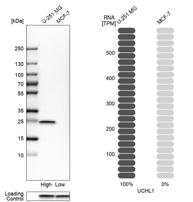
- Main image

- Experimental details
- Western blot analysis in human cell lines U-251MG and MCF-7 using Anti-UCHL1 antibody. Corresponding UCHL1 RNA-seq data are presented for the same cell lines. Loading control: Anti-GAPDH.
Supportive validation
- Submitted by
- Atlas Antibodies (provider)
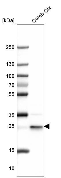
- Main image
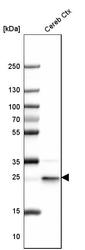
- Experimental details
- Western blot analysis in mouse cerebral cortex tissue.
- Submitted by
- Atlas Antibodies (provider)
- Main image

- Experimental details
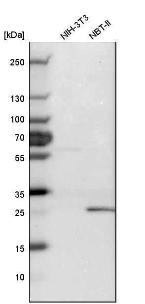
- Western blot analysis in mouse cell line NIH-3T3 and rat cell line NBT-II.
Supportive validation
- Submitted by
- Atlas Antibodies (provider)
- Main image

- Experimental details
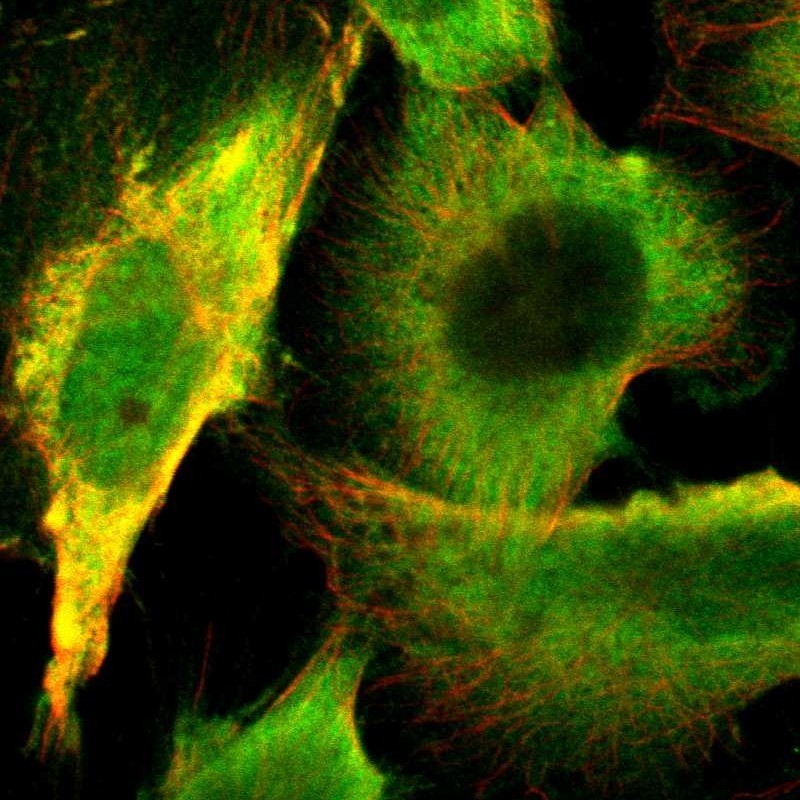
- Immunofluorescent staining of human cell line U-251 MG shows localization to nucleoplasm & cytosol.
- Sample type
- HUMAN
Enhanced validation
Supportive validation
- Submitted by
- Atlas Antibodies (provider)
- Enhanced method
- Orthogonal validation
- Main image
- Experimental details
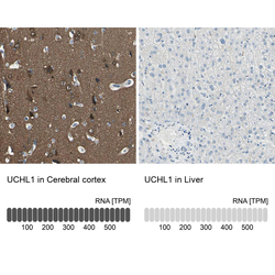
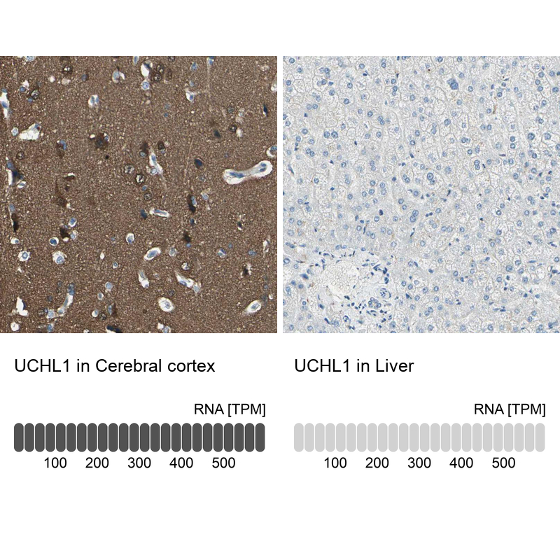
- Immunohistochemistry analysis in human cerebral cortex and liver tissues using HPA005993 antibody. Corresponding UCHL1 RNA-seq data are presented for the same tissues.
- Sample type
- HUMAN
Supportive validation
- Submitted by
- Atlas Antibodies (provider)
- Main image
- Experimental details
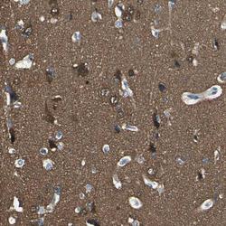
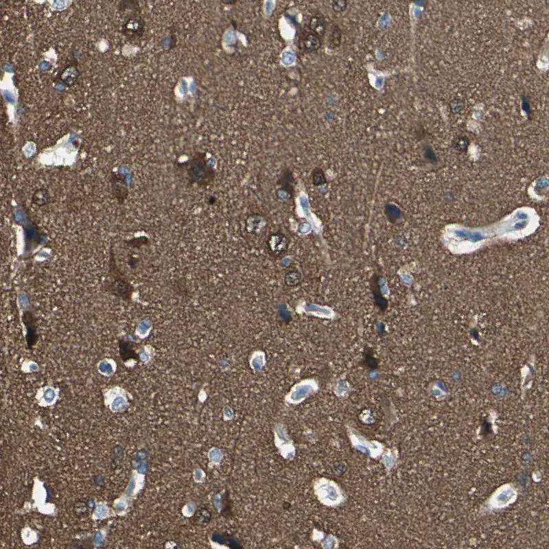
- Immunohistochemical staining of human cerebral cortex shows strong cytoplasmic positivity in neurons and in neuropil.
- Sample type
- HUMAN
- Submitted by
- Atlas Antibodies (provider)
- Main image
- Experimental details
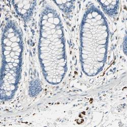
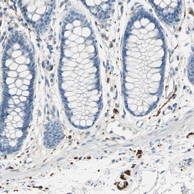
- Immunohistochemical staining of human rectum shows strong cytoplasmic positivity in peripheral ganglion and peripheral nerves.
- Sample type
- HUMAN
- Submitted by
- Atlas Antibodies (provider)
- Main image
- Experimental details
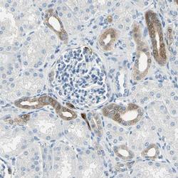
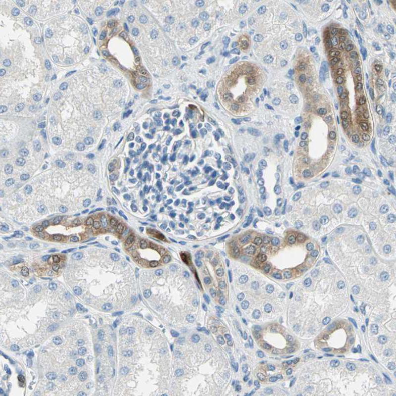
- Immunohistochemical staining of human kidney shows moderate cytoplasmic positivity in cells in tubules.
- Sample type
- HUMAN
- Submitted by
- Atlas Antibodies (provider)
- Main image
- Experimental details
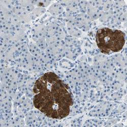
- Immunohistochemical staining of human pancreas shows strong cytoplasmic positivity in islets of Langerhans.
- Sample type
- HUMAN

- Submitted by
- Atlas Antibodies (provider)
- Main image
- Experimental details
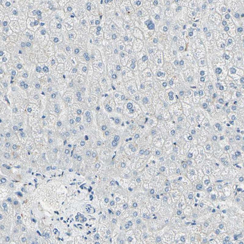
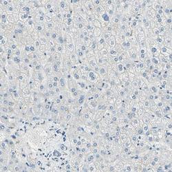
- Immunohistochemical staining of human liver shows no positivity in hepatocytes, as expected.
- Sample type
- HUMAN