 Explore
 Validate
 Learn
LearnMA5-15077
antibody from Invitrogen Antibodies
Targeting: BIRC5
API4, EPR-1, survivin
 Western blot Immunocytochemistry
 Immunoprecipitation Immunohistochemistry Flow cytometry Other assay
Antibody data
- Antibody Data
- Antigen structure
- References [3]
- Comments [0]
- Validations
- Western blot [2]
- Immunocytochemistry [2]
- Immunohistochemistry [1]
- Flow cytometry [1]
- Other assay [3]
Submit
Validation data
Reference
Comment
Report error
- Product number
- MA5-15077 - Provider product page

- Provider
- Invitrogen Antibodies
- Product name
- Survivin Monoclonal Antibody (J.33.5)
- Antibody type
- Monoclonal
- Antigen
- Synthetic peptide
- Description
- It is not recommended to aliquot this antibody.
- Reactivity
- Human, Mouse, Rat
- Host
- Rabbit
- Isotype
- IgG
- Antibody clone number
- J.33.5
- Vial size
- 100 µL
- Concentration
- 311 µg/mL
- Storage
- -20°C
Submitted references Development of Multi-Scale X-ray Fluorescence Tomography for Examination of Nanocomposite-Treated Biological Samples.
Therapeutic potential of cannabidiol against ischemia/reperfusion liver injury in rats.
Expression of survivin, CD117, and C-erbB-2 in neuroendocrine carcinoma of the uterine cervix.
Chen S, Lastra RO, Paunesku T, Antipova O, Li L, Deng J, Luo Y, Wanzer MB, Popovic J, Li Y, Glasco AD, Jacobsen C, Vogt S, Woloschak GE
Cancers 2021 Sep 6;13(17)
Cancers 2021 Sep 6;13(17)
Therapeutic potential of cannabidiol against ischemia/reperfusion liver injury in rats.
Fouad AA, Jresat I
European journal of pharmacology 2011 Nov 16;670(1):216-23
European journal of pharmacology 2011 Nov 16;670(1):216-23
Expression of survivin, CD117, and C-erbB-2 in neuroendocrine carcinoma of the uterine cervix.
Sukpan K, Settakorn J, Khunamornpong S, Cheewakriangkrai C, Srisomboon J, Siriaunkgul S
International journal of gynecological cancer : official journal of the International Gynecological Cancer Society 2011 Jul;21(5):911-7
International journal of gynecological cancer : official journal of the International Gynecological Cancer Society 2011 Jul;21(5):911-7
No comments: Submit comment
Supportive validation
- Submitted by
- Invitrogen Antibodies (provider)
- Main image
- Experimental details
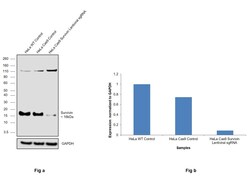
- CRISPR-Cas9 mediated genome editing ofSurvivin (as confirmed by next generation sequencing) was achieved by using LentiArray™ Lentiviral sgRNA (Product # A32042, AssayID CRISPR711998_LV) and LentiArray Cas9 Lentivirus (Product # A32064). Fig (a) Western blot analysis of Survivin was performed by loading 30 µg of HeLa Wild Type (Lane 1), HeLa Cas9 (Lane 2) and HeLa Cas9 cells transduced with Survivin Lentiviral sgRNA (Lane 3) whole cell extracts. The samples were electrophoresed using NuPAGE™ Novex™ 4-12% Bis-Tris Protein Gel (Product # NP0322BOX). Resolved proteins were then transferred onto a nitrocellulose membrane (Product # IB23001) by iBlot® 2 Dry Blotting System (Product # IB21001). The blot was probed with Anti-Survivin Monoclonal Antibody (J.33.5) (Product # MA5-15077) using 1:1,000 dilution and Goat anti-Rabbit IgG (H+L) Superclonal™ Recombinant Secondary Antibody, HRP (Product # A27036 1:5,000 dilution).Chemiluminescent detection was performed using Novex® ECL Chemiluminescent Substrate Reagent Kit (Product # WP20005). A reduced signal in sgRNA transduced cells using the LentiArray™ CRISPR product line confirms that antibody is specific toSurvivin (Fig (b)). An uncharacterized band was observed in all the samples at ~110 kDa.

- Submitted by
- Invitrogen Antibodies (provider)
- Main image
- Experimental details
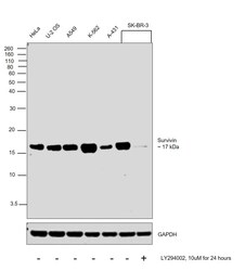
- Western blot was performed using Anti-Survivin Monoclonal Antibody (J.33.5) (Product # MA5-15077) and a 17 kDa band corresponding to Survivin was observed across cell lines tested and was lost upon LY294002 treatment in SK-BR-3. Whole cell extracts (30 µg lysate) of HeLa (Lane 1), U-2 OS (Lane 2), A549 (Lane 3), K-562 (Lane 4), A-431 (Lane 5), SK-BR-3 (Lane 6) and SK-BR-3 treated with LY294002 (10uM for 24 hours) (Lane 7) were electrophoresed using NuPAGE™ 10% Bis-Tris Protein Gel (Product # NP0302BOX). Resolved proteins were then transferred onto a Nitrocellulose membrane (Product # IB23001) by iBlot® 2 Dry Blotting System (Product # IB21001). The blot was probed with the primary antibody (1:1000 dilution) and detected by chemiluminescence with Goat anti-Rabbit IgG (H+L) Superclonal™ Recombinant Secondary Antibody, HRP (Product # A27036, 1:4000 dilution) using the iBright FL 1000 (Product # A32752). Chemiluminescent detection was performed using Novex® ECL Chemiluminescent Substrate Reagent Kit (Product # WP20005).
Supportive validation
- Submitted by
- Invitrogen Antibodies (provider)
- Main image

- Experimental details
- Immunofluorescent analysis of Survivin in HeLa cells using a Survivin monoclonal antibody (Product # MA5-15077) (green). Mitochondria have been labeled with a mitochondrial fluorescent red dye. DNA is labeled using a fluorescent blue dye.
- Submitted by
- Invitrogen Antibodies (provider)
- Main image
- Experimental details
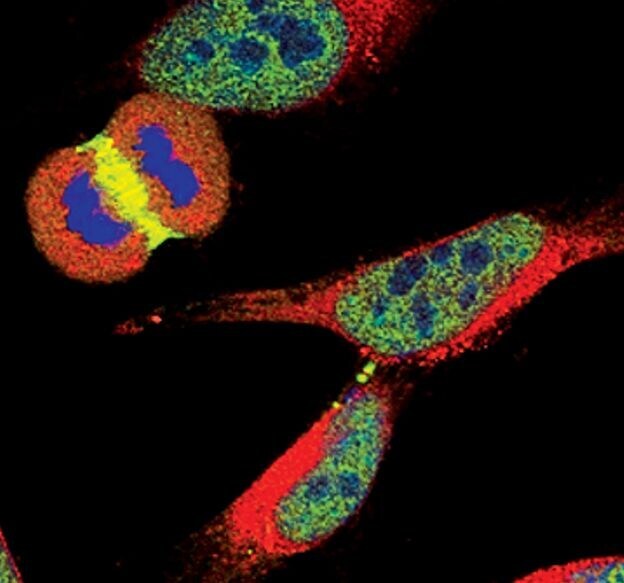
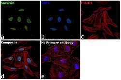
- Immunofluorescence analysis of Survivin was performed using 70% confluent log phase HeLa cells. The cells were fixed with 4% paraformaldehyde for 10 minutes, permeabilized with 0.1% Triton™ X-100 for 15 minutes, and blocked with 2% BSA for 45 minutes at room temperature. The cells were labeled with Survivin Monoclonal Antibody (J.33.5) (Product # MA5-15077) at 1:500 dilution in 0.1% BSA, incubated at 4 degree celsius overnight and then labeled with Donkey anti-Rabbit IgG (H+L) Highly Cross-Adsorbed Secondary Antibody, Alexa Fluor Plus 488 (Product # A32790), (1:2000 dilution), for 45 minutes at room temperature (Panel a: Green). Nuclei (Panel b:Blue) were stained with ProLong™ Diamond Antifade Mountant with DAPI (Product # P36962). F-actin (Panel c: Red) was stained with Rhodamine Phalloidin (Product # R415, 1:300). Panel d represents the merged image showing Nuclear localization. Panel e represents control cells with no primary antibody to assess background. The images were captured at 60X magnification.
Supportive validation
- Submitted by
- Invitrogen Antibodies (provider)
- Main image
- Experimental details
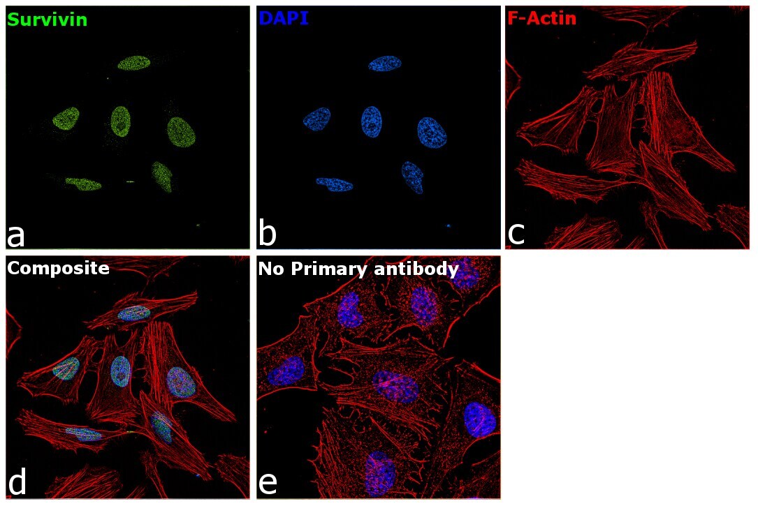
- Immunohistochemical analysis of Survivin in paraffin-embedded human lung carcinoma using a Survivin monoclonal antibody (Product # MA5-15077) showing nuclear localization.

Supportive validation
- Submitted by
- Invitrogen Antibodies (provider)
- Main image
- Experimental details
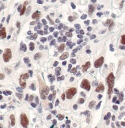
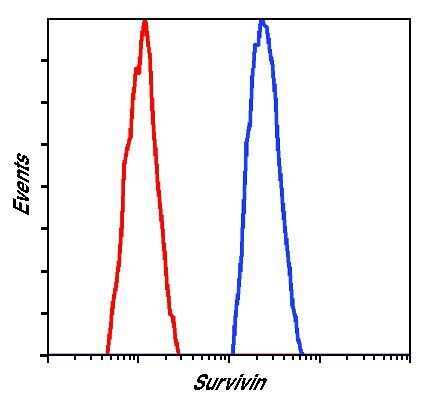
- Flow cytometric analysis of Survivin in untreated Jurkat cells using a Survivin monoclonal antibody (Product # MA5-15077) (blue) compared to a nonspecific negative control antibody (red).
Supportive validation
- Submitted by
- Invitrogen Antibodies (provider)
- Main image

- Experimental details
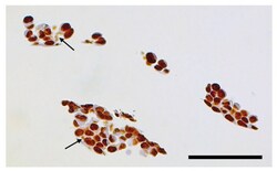
- Figure 7 Immunohistochemistry for BIRC5 shows typically stained cell nuclei (for a control tissue sample see Figure S2 ). However, in addition to cell nuclei, some lightly stained material is also noticeable in the cytosol of many cells (e.g., areas indicated by arrows), possibly containing aggregates of nanocomposites with BIRC5 attached to particle surface in a manner permitting its recognition by the antibodies. Black bar corresponds to 100 microns.

- Submitted by
- Invitrogen Antibodies (provider)
- Main image

- Experimental details
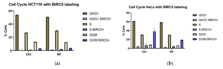
- Figure 2 Cell cycle distribution of control and nanocomposite treated cells with BIRC5 labelling. ( a ) Distribution for control and nanocomposite treated HCT116 cells. ( b ) Distribution for control and nanocomposite treated HeLa cells. Analysis for the entire population of cells (>=50,000 per biological replicate) is shown in the brown bars. Purple colored bars, as indicated in the figure legend, apply to BIRC5 positive cells: the most intensely stained ~1000 cells in each case. Each bar represents the average and standard deviation from triplicate experiments; for gating strategy, see Figure S3 ; for cell numbers, BIRC5 staining intensity, and other details, see Table S1 .
- Submitted by
- Invitrogen Antibodies (provider)
- Main image
- Experimental details
- Figure 7 Immunohistochemistry for BIRC5 shows typically stained cell nuclei (for a control tissue sample see Figure S2 ). However, in addition to cell nuclei, some lightly stained material is also noticeable in the cytosol of many cells (e.g., areas indicated by arrows), possibly containing aggregates of nanocomposites with BIRC5 attached to particle surface in a manner permitting its recognition by the antibodies. Black bar corresponds to 100 microns.
