 Explore
 Validate
 Learn
LearnPA5-102743
antibody from Invitrogen Antibodies
Targeting: LTBR
D12S370, TNF-R-III, TNFCR, TNFR-RP, TNFR2-RP, TNFRSF3
 Western blot
Antibody data
- Antibody Data
- Antigen structure
- References [0]
- Comments [0]
- Validations
- Western blot [2]
- Immunocytochemistry [1]
- Immunohistochemistry [1]
Submit
Validation data
Reference
Comment
Report error
- Product number
- PA5-102743 - Provider product page

- Provider
- Invitrogen Antibodies
- Product name
- LTBR Polyclonal Antibody
- Antibody type
- Polyclonal
- Antigen
- Synthetic peptide
- Reactivity
- Human, Mouse
- Host
- Rabbit
- Isotype
- IgG
- Vial size
- 100 µL
- Concentration
- 1 mg/mL
- Storage
- -20°C
No comments: Submit comment
Supportive validation
- Submitted by
- Invitrogen Antibodies (provider)
- Main image

- Experimental details
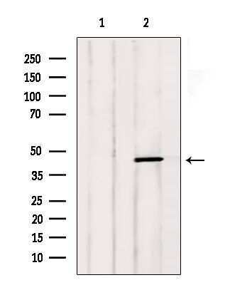
- Western blot analysis of LTBR in mouse cancer (Lane 1: treated with blocking peptide). Samples were incubated with LTBR polyclonal antibody (Product # PA5-102743).
- Submitted by
- Invitrogen Antibodies (provider)
- Main image

- Experimental details
- Western Blot was performed using Anti-LTBR Polyclonal Antibody (Product # PA5-102743) and a 55 kDa band corresponding to Tumor necrosis factor receptor superfamily member 3 was observed in HeLa and A549 which are reported to be positive cell lines and not in SH-SY5Y and HEK-293 which are reported to be negative. Membrane enriched extracts (30 µg lysate) of HeLa (Lane 1), A549 (Lane 2), SH-SY5Y (Lane 3) and HEK-293 (Lane 4) were electrophoresed using NuPAGE™ 10% Bis-Tris Protein Gel (Product # NP0301BOX). Resolved proteins were then transferred onto a nitrocellulose membrane (Product # IB23001) by iBlot® 2 Dry Blotting System (Product # IB21001). The blot was probed with the primary antibody (1:1000 dilution) and detected by chemiluminescence with Goat anti-Rabbit IgG (H+L) Superclonal™ Recombinant Secondary Antibody, HRP (Product # A27036, 1:4000 dilution) using the iBright FL 1000 (Product # A32752). Chemiluminescent detection was performed using SuperSignal™ West Dura Extended Duration Substrate (Product # 34076). An uncharacterized band of ~48 kDa was also observed across the cell lines tested.

Supportive validation
- Submitted by
- Invitrogen Antibodies (provider)
- Main image

- Experimental details
- Immunofluorescent analysis of LTBR in HeLa cells. Samples were fixed with paraformaldehyde, permeabilized with 0.1% Triton X-100, blocked with 10% serum (45 min at 25°C) incubated with LTBR polyclonal antibody (Product # PA5-102743) using a dilution of 1:200 (1 hr, 37°C), and followed by goat anti-rabbit IgG Alexa Fluor 594 at a dilution of 1:600.

Supportive validation
- Submitted by
- Invitrogen Antibodies (provider)
- Main image
- Experimental details
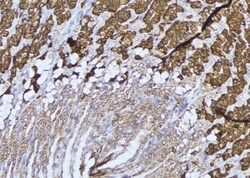
- Immunohistochemistry analysis of paraffin-embedded LTBR in human gastric tissue. Antigen retrieval was performed using citrate buffer. Samples were blocked with blocking buffer (1.5 hr, 22°C), incubated with LTBR polyclonal antibody (Product # PA5-102743) using a dilution of 1:100 (1.5 hr, 22°C), followed by HRP conjugated goat anti-rabbit.
