Explore
 Validate
 Learn
Learn Western blot
Antibody data
- Antibody Data
- Antigen structure
- References [2]
- Comments [0]
- Validations
- Western blot [1]
- Immunocytochemistry [2]
- Immunohistochemistry [3]
- Other assay [4]
Submit
Validation data
Reference
Comment
Report error
- Product number
- PA5-87536 - Provider product page

- Provider
- Invitrogen Antibodies
- Product name
- Caspase 1 Polyclonal Antibody
- Antibody type
- Polyclonal
- Antigen
- Synthetic peptide
- Description
- Immunogen sequence: MADKVLKEKR KLFIRSMGEA PQAVQDNPAM PTSSGSEGNV KLCSLEEAQR IWKQKSAEIY PIMDKSSRTR LALIICNEEF DSIPRRTGAE VDITGMTMLL QNLGYSVDVK KNLTASDMTT ELEAFAHRPE HKTSDSTFLV FMSHGIREGI CGKKHSEQVP DILQLNAIFN MLNTKNCPSL KDKPKVIIIQ ACRGDSPGVV WFKDSVGVSG NLSLPTTEEF EDDAIKKAHI EKDFIAFCSS TPDNVSWRHP TMGSVFIGRL IEHMQEYACS CDVEEIFRKV RFSFEQPDGR AQMPTTERVT LTRCFYLFPG H; Positive Samples: THP-1, Mouse lung, Rat spleen; Cellular Location: Cytoplasm
- Reactivity
- Human, Mouse
- Host
- Rabbit
- Isotype
- IgG
- Vial size
- 100 µL
- Concentration
- 0.55 mg/mL
- Storage
- -20° C, Avoid Freeze/Thaw Cycles
Submitted references IP-Se-06, a Selenylated Imidazo[1,2-a]pyridine, Modulates Intracellular Redox State and Causes Akt/mTOR/HIF-1α and MAPK Signaling Inhibition, Promoting Antiproliferative Effect and Apoptosis in Glioblastoma Cells.
Modulation of Gut Microbiota Combined with Upregulation of Intestinal Tight Junction Explains Anti-Inflammatory Effect of Corylin on Colitis-Associated Cancer in Mice.
Dos Santos DC, Rafique J, Saba S, Grinevicius VMAS, Filho DW, Zamoner A, Braga AL, Pedrosa RC, Ourique F
Oxidative medicine and cellular longevity 2022;2022:3710449
Oxidative medicine and cellular longevity 2022;2022:3710449
Modulation of Gut Microbiota Combined with Upregulation of Intestinal Tight Junction Explains Anti-Inflammatory Effect of Corylin on Colitis-Associated Cancer in Mice.
Chang ZY, Liu HM, Leu YL, Hsu CH, Lee TY
International journal of molecular sciences 2022 Feb 28;23(5)
International journal of molecular sciences 2022 Feb 28;23(5)
No comments: Submit comment
Supportive validation
- Submitted by
- Invitrogen Antibodies (provider)
- Main image
- Experimental details
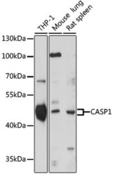
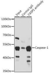
- Western blot analysis of extracts of various cell lines, using CASP1 Polyclonal antibody (Product # PA5-87536) at 1:1000 dilution. Secondary antibody: HRP Goat Anti-Rabbit IgG (H+L) at 1:10000 dilution. Lysates/proteins: 25ug per lane. Blocking buffer: 3% nonfat dry milk in TBST. Exposure time: 30s.

Supportive validation
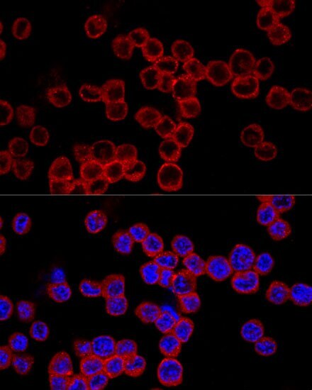
- Submitted by
- Invitrogen Antibodies (provider)
- Main image
- Experimental details
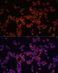
- Immunocytochemistry-Immunofluorescence analysis of Caspase 1 was performed in Raw264.7 cells using Caspase 1 Polyclonal Antibody (Product # PA5-87536) at a dilution of 1:100. Blue: DAPI for nuclear staining.
- Submitted by
- Invitrogen Antibodies (provider)
- Main image
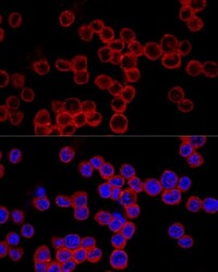
- Experimental details
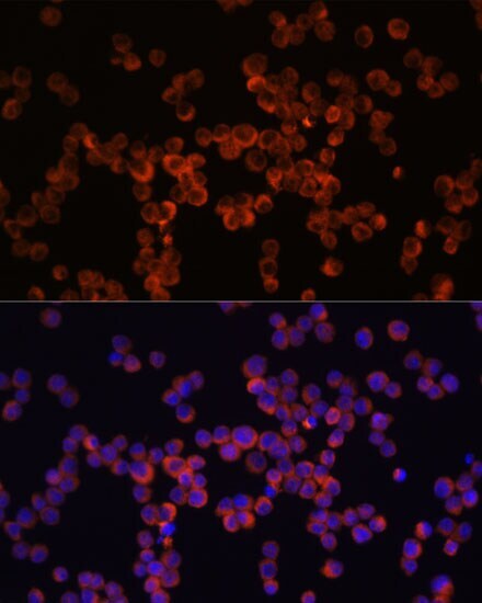
- Immunocytochemistry-Immunofluorescence analysis of Caspase 1 was performed in Raw264.7 cells using Caspase 1 Polyclonal Antibody (Product # PA5-87536) at a dilution of 1:200. Blue: DAPI for nuclear staining.
Supportive validation
- Submitted by
- Invitrogen Antibodies (provider)
- Main image
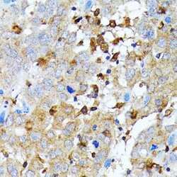
- Experimental details
- Immunohistochemistry analysis of Caspase 1 in paraffin-embedded human liver cancer using Caspase 1 Polyclonal Antibody (Product # PA5-87536) at a dilution of 1:100.

- Submitted by
- Invitrogen Antibodies (provider)
- Main image
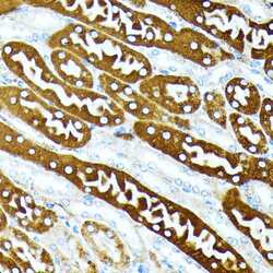
- Experimental details
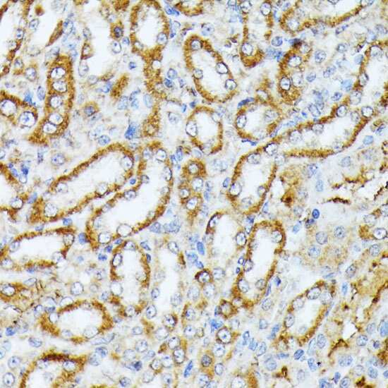
- Immunohistochemistry analysis of Caspase 1 in paraffin-embedded mouse kidney using Caspase 1 Polyclonal Antibody (Product # PA5-87536) at a dilution of 1:100.

- Submitted by
- Invitrogen Antibodies (provider)
- Main image
- Experimental details
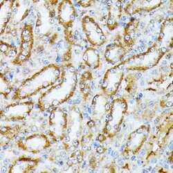
- Immunohistochemistry analysis of Caspase 1 in paraffin-embedded rat kidney using Caspase 1 Polyclonal Antibody (Product # PA5-87536) at a dilution of 1:100.
Supportive validation
- Submitted by
- Invitrogen Antibodies (provider)
- Main image
- Experimental details
- Immunoprecipitation analysis of Caspase 1 was performed in 200 µg extracts of THP-1 cells using Caspase 1 Polyclonal Antibody (Product # PA5-87536). Western blot was performed from the immunoprecipitate using Caspase 1 Polyclonal Antibody.

- Submitted by
- Invitrogen Antibodies (provider)
- Main image
- Experimental details
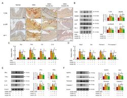
- Corylin improves the TLR4 signal pathway in DSS-induced colitis mice. ( A ) Expression analysis by immunohistochemical staining of TLR4, p-p38, and AP-1 in mice after the indicated treatment. Scale bar: 100 mum. Red arrows highlight positive staining. ( B ) Western blot analysis of TLR4, MyD88, p-p38, AP-1, and beta-actin (loading control) in colon homogenates. Right graph indicates quantification relative to beta-actin. ( C , D ) mRNA expression of Ifngamma , Tnf-alpha , Il-6 , Il-1beta , Nlrp3 , Asc , Pannexin , and Pro-caspase 1 determined by qRT-PCR. ( E , F ) Western blot analysis of IFNgamma, TNF-alpha, IL-6, IL-1beta, NLRP3, ASC, Pannexin, and Caspase 1, and beta-actin (loading control) in colon homogenates. Results represent mean +- SEM. * p < 0.05, Normal compared with DSS; # p < 0.05, DSS compared with DSS + Corylin (L); Y= p < 0.05, DSS compared with DSS + Corylin (H); SS p < 0.05, DSS + Corylin (L) compared with DSS + Corylin (H). DSS, dextran sodium sulfate; TLR4, Toll-like receptor 4; MyD88, myeloid differentiation primary response 88; AP-1, activator protein 1; Ifngamma , interferon gamma; Tnf-alpha , tumor necrosis factor-alpha; Il-6 , interleukin-6; Nlrp3 , NACHT, LRR, and PYD domains-containing protein 3.

- Submitted by
- Invitrogen Antibodies (provider)
- Main image
- Experimental details
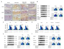
- Corylin improves the TLR4 signaling pathway in AOM/DSS-induced-colitis-associated colorectal cancer mice. ( A ) Expression analysis by immunohistochemical staining of TLR4, p-p38, and AP-1 in mice after the indicated treatment. Scale bar: 100 mum. ( B ) Western blot analysis of TLR4, MyD88, p-p38, AP-1, and beta-actin (loading control) in colon. ( C , D ) mRNA expression of Ifngamma , Tnf-alpha , Il-6 , Il-1beta , Nlrp3 , Asc , Pannexin , and Pro-caspase 1 as determined by qRT-PCR. ( E , F ) Western blot analysis of IFNgamma, TNF-alpha, IL-6, IL-1beta, NLRP3, ASC, Pannexin, and Caspase 1, and beta-actin (loading control) in colon homogenates. Results represent mean +- SEM. * p < 0.05, Normal compared with AOM/DSS; # p < 0.05, AOM/DSS compared with AOM/DSS + Corylin (L); Y= p < 0.05, AOM/DSS compared with AOM/DSS + Corylin (H); SS p < 0.05, DSS + Corylin (L) compared with DSS + Corylin (H). AOM, azoxymethane; DSS, dextran sodium sulfate; TLR4, Toll-like receptor 4; MyD88, myeloid differentiation primary response 88; AP-1, activator protein 1; Ifngamma , interferon gamma; Tnf-alpha , tumor necrosis factor-alpha; Il-6 , interleukin-6; Nlrp3 , NACHT, LRR, and PYD domains-containing protein 3.

- Submitted by
- Invitrogen Antibodies (provider)
- Main image
- Experimental details
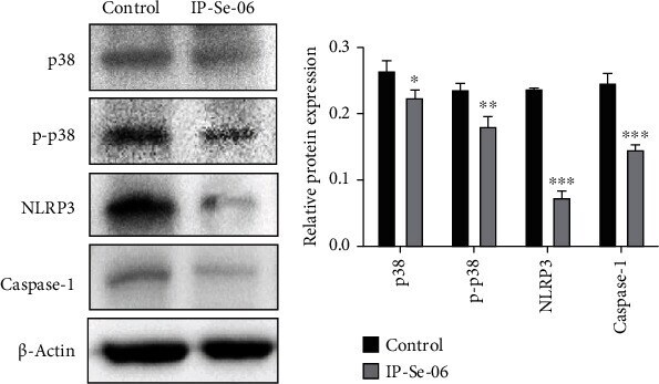
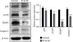
- Treatment with IP-Se-06 (1 mu M) at 48 h downregulated p38 mitogen-activated protein kinase and inflammasome complex protein in A172 glioblastoma cells. (a) Immunoblotting analysis detected the downregulation of p38, p-p38, NLRP3 and caspase-1 protein levels. beta -Actin was used as a loading control. (b) Densitometric analysis results of p38, p-p38, NLRP3 and caspase-1 were quantified with ImageJ. The data were considered as statistically significant at * p < 0.05, ** p < 0.01, and *** p < 0.001 compared to the control group (nontreated cells).