Antibody data
- Antibody Data
- Antigen structure
- References [4]
- Comments [0]
- Validations
- Western blot [2]
- Immunocytochemistry [1]
- Immunohistochemistry [2]
- Other assay [1]
Submit
Validation data
Reference
Comment
Report error
- Product number
- MA1-06104 - Provider product page

- Provider
- Invitrogen Antibodies
- Product name
- Lamin B2 Monoclonal Antibody (LN43)
- Antibody type
- Monoclonal
- Antigen
- Other
- Description
- MA1-06104 detects lamin B2 in human, mouse, porcine, Xenopus laevis, zebrafish and hamster samples. MA1-06104 has sucessfully been used in FACS, immunohistochemistry, and Western blotting procedures. The MA1-06104 immunogen is a detergent insoluble fraction of potoroo cell line PtK1. Store at 4ºC, or in small aliquots at -20ºC.
- Reactivity
- Human, Mouse, Hamster, Porcine, Xenopus, Zebrafish
- Host
- Mouse
- Isotype
- IgG
- Antibody clone number
- LN43
- Vial size
- 100 µL
- Concentration
- 1 mg/mL
- Storage
- Store at 4°C short term. For long term storage, store at -20°C, avoiding freeze/thaw cycles.
Submitted references Antioxidant Activity of Valeriana fauriei Protects against Dexamethasone-Induced Muscle Atrophy.
Transcription upregulation via force-induced direct stretching of chromatin.
Inhibition of the transient receptor potential melastatin-2 channel causes increased DNA damage and decreased proliferation in breast adenocarcinoma cells.
Activation of cell death mediated by apoptosis-inducing factor due to the absence of poly(ADP-ribose) glycohydrolase.
Kim YI, Lee H, Nirmala FS, Seo HD, Ha TY, Jung CH, Ahn J
Oxidative medicine and cellular longevity 2022;2022:3645431
Oxidative medicine and cellular longevity 2022;2022:3645431
Transcription upregulation via force-induced direct stretching of chromatin.
Tajik A, Zhang Y, Wei F, Sun J, Jia Q, Zhou W, Singh R, Khanna N, Belmont AS, Wang N
Nature materials 2016 Dec;15(12):1287-1296
Nature materials 2016 Dec;15(12):1287-1296
Inhibition of the transient receptor potential melastatin-2 channel causes increased DNA damage and decreased proliferation in breast adenocarcinoma cells.
Hopkins MM, Feng X, Liu M, Parker LP, Koh DW
International journal of oncology 2015 May;46(5):2267-76
International journal of oncology 2015 May;46(5):2267-76
Activation of cell death mediated by apoptosis-inducing factor due to the absence of poly(ADP-ribose) glycohydrolase.
Zhou Y, Feng X, Koh DW
Biochemistry 2011 Apr 12;50(14):2850-9
Biochemistry 2011 Apr 12;50(14):2850-9
No comments: Submit comment
Supportive validation
- Submitted by
- Invitrogen Antibodies (provider)
- Main image

- Experimental details
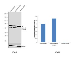
- Knockdown of Lamin B2 was achieved by transfecting HCT 116 with Lamin B2 specific siRNAs (Silencer® select Product # s39477, Product # s39478). Western blot analysis (Fig. a) was performed using whole cell extracts from the Lamin B2 knockdown cells (Lane 3), non-specific scrambled siRNA transfected cells (Lane 2) and untransfected cells (Lane 1). The blot was probed with Lamin B2 Monoclonal Antibody (Product # MA1-06104, 1:1000 dilution) and Goat anti-Mouse IgG (H+L), Superclonal™ Recombinant Secondary Antibody, HRP conjugate (Product # A28177, 1:4000 dilution). Densitometric analysis of this western blot is shown in histogram (Fig. b). Decrease in signal upon siRNA mediated knock down confirms that antibody is specific to Lamin B2. An uncharacterized band (*) at ~75 kDa was also observed.
- Submitted by
- Invitrogen Antibodies (provider)
- Main image

- Experimental details
- Western blot was performed using Anti-Lamin B2 Monoclonal Antibody (LN43) (Product # MA1-06104) and a 70 kDa band corresponding to Lamin B2 along with uncharacterized bands (*) at ~50-55 kDa was observed in the cell lines tested. Modified whole cell extracts (1%SDS) (30 µg lysate) of Jurkat (Lane 1), HCT 116 (Lane 2), U-87 MG (Lane 3), IMR32 (Lane 4), Hep G2 (Lane 5), HeLa (Lane 6) and MDA-MB-231 (Lane 7) were electrophoresed using Novex® NuPAGE® 4-12 % Bis-Tris gel (Product # NP0321BOX). Resolved proteins were then transferred onto a nitrocellulose membrane (Product # IB23001) by iBlot® 2 Dry Blotting System (Product # IB21001). The blot was probed with the primary antibody (1:1000 dilution) and detected by chemiluminescence with Goat anti-Mouse IgG (H+L), Superclonal™ Recombinant Secondary Antibody, HRP conjugate (Product # A28177, 1:4000 dilution) using the iBright FL 1000 (Product # A32752). Chemiluminescent detection was performed using Novex® ECL Chemiluminescent Substrate Reagent Kit (Product # WP20005).
Supportive validation
- Submitted by
- Invitrogen Antibodies (provider)
- Main image

- Experimental details
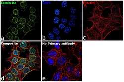
- Immunofluorescence analysis of Lamin B2 was performed using 70% confluent log phase HCT 116 cells. The cells were fixed with 4% paraformaldehyde for 10 minutes, permeabilized with 0.1% Triton™ X-100 for 15 minutes, and blocked with 2% BSA for 1 hour at room temperature. The cells were labeled with Lamin B2 Monoclonal Antibody (LN43) (Product # MA1-06104) at 1:200 dilution in 0.1% BSA, incubated at 4 degree Celsius overnight and then with Goat anti-Mouse IgG (H+L), Superclonal™ Recombinant Secondary Antibody, Alexa Fluor 488 conjugate (Product # A28175) at a dilution of 1:2000 for 45 minutes at room temperature (Panel a: green). Nuclei (Panel b: blue) were stained with SlowFade® Gold Antifade Mountant with DAPI (Product # S36938). F-actin (Panel c: red) was stained with Rhodamine Phalloidin (Product # R415, 1:300). Panel d represents the merged image showing staining in nuclear periphery. Panel e represents control cells with no primary antibody to assess background. The images were captured at 60X magnification.
Supportive validation
- Submitted by
- Invitrogen Antibodies (provider)
- Main image

- Experimental details
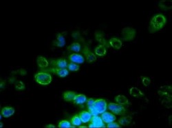
- Immunofluorescent analysis of 9 days old zebrafish embryo using Lamin B2 monoclonal antibody (Product # MA1-06104).
- Submitted by
- Invitrogen Antibodies (provider)
- Main image

- Experimental details
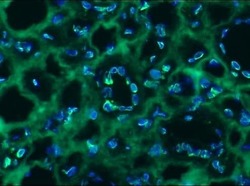
- Immunohistochemistry on frozen sections of human kidney showing nuclear lamina staining in the ductal epithelium stained with Lamin B2 monoclonal antibody (Product # MA1-06104).
Supportive validation
- Submitted by
- Invitrogen Antibodies (provider)
- Main image

- Experimental details
- Figure 4 Nuclear localization of TRPM2 in breast cancer cells. (A) Noncancerous breast cell lines, HMEC and MCF-10A (a), and cancerous breast cell lines, MCF-7 and MDA-MB-231 (b), were fractionated into subcellular fractions (NE-PER Extraction kit, Pierce) and cellular localization of TRPM2 was then analyzed by immunoblot. Immunoblot detection of mitochondrial manganese superoxide dismutase (MnSOD) and nuclear lamin B2 was performed to verify successful fractionations. (B) Quantification of protein levels of TRPM2 in the cytoplasmic (Cyto) and nuclear (Nuc) fractions of MCF-7 and MDA-MB-231 cells by densitometry. Quantification is based on three immunoblots. Error bars represent SEM.
 Explore
 Validate
 Learn
Learn Western blot
 Flow cytometry