Explore
 Validate
 Learn
LearnAF5578
antibody from R&D Systems
Targeting: S100A9
60B8AG, CAGB, CFAG, CGLB, LIAG, MAC387, MIF, MRP14, NIF, P14
 Western blot
Antibody data
- Antibody Data
- Antigen structure
- References [0]
- Comments [0]
- Validations
- Western blot [2]
- Immunocytochemistry [2]
- Immunohistochemistry [1]
- Flow cytometry [1]
Submit
Validation data
Reference
Comment
Report error
- Product number
- AF5578 - Provider product page

- Provider
- R&D Systems
- Product name
- Human S100A9 Antibody
- Antibody type
- Polyclonal
- Description
- Antigen Affinity-purified. Detects human S100A9 in direct ELISAs and Western blots. In direct ELISAs, less than 5% cross-reactivity with recombinant mouse S100A9 is observed.
- Reactivity
- Human
- Host
- Sheep
- Conjugate
- Unconjugated
- Antigen sequence
P06702- Isotype
- IgG
- Vial size
- 100 ug
- Concentration
- LYOPH
- Storage
- Use a manual defrost freezer and avoid repeated freeze-thaw cycles. 12 months from date of receipt, -20 to -70 °C as supplied. 1 month, 2 to 8 °C under sterile conditions after reconstitution. 6 months, -20 to -70 °C under sterile conditions after reconstitution.
No comments: Submit comment
Supportive validation
- Submitted by
- R&D Systems (provider)
- Main image

- Experimental details
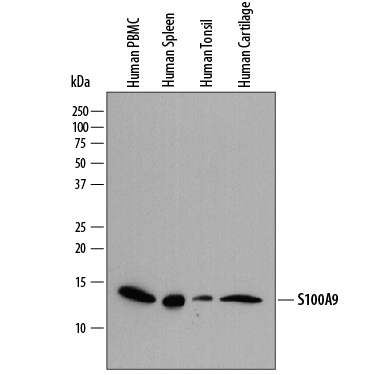
- Detection of Human S100A9 by Western Blot. Western blot shows lysates of human peripheral blood mononuclear cells (PBMC), human spleen tissue, human tonsil tissue, and human cartilage tissue. PVDF membrane was probed with 0.2 µg/mL of Sheep Anti-Human S100A9 Antigen Affinity-purified Polyclonal Antibody (Catalog # AF5578) followed by HRP-conjugated Anti-Sheep IgG Secondary Antibody (Catalog # HAF016). A specific band was detected for S100A9 at approximately 14 kDa (as indicated). This experiment was conducted under reducing conditions and using Immunoblot Buffer Group 1.
- Submitted by
- R&D Systems (provider)
- Main image

- Experimental details
- Western Blot Shows Human S100A9 Specificity by Using Knockout Cell Line. Western blot shows lysates of MDA-MB-468 human breast cancer parental cell line and S100A9 knock out MDA-MB-468 cell line (KO). PVDF membrane was probed with 0.2 µg/mL of Sheep Anti-Human S100A9 Antigen Affinity-purified Polyclonal Antibody (Catalog # AF5578) followed by HRP-conjugated Anti-Sheep IgG Secondary Antibody (Catalog # HAF016). A specific band was detected for S100A9 at approximately 14 kDa (as indicated) in the parental MDA-MB-468 cell line, but is not detectable in knockout MDA-MB-468 cell line. This experiment was conducted under reducing conditions and using Immunoblot Buffer Group 1.

Supportive validation
- Submitted by
- R&D Systems (provider)
- Main image
- Experimental details
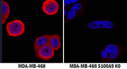
- S100A9 Specificity is Shown by Immunocytochemistry in Knockout Cell Line. S100A9 was detected in immersion fixed MDA-MB-468 human breast cancer cell line but is not detected in S100A9 knockout (KO) MDA-MB-468 cell line using Sheep Anti-Human S100A9 Antigen Affinity-purified Polyclonal Antibody (Catalog # AF5578) at 1.7 µg/mL for 3 hours at room temperature. Cells were stained using the NorthernLights 557-conjugated Anti-Sheep IgG Secondary Antibody (red; Catalog # NL010) and counterstained with DAPI (blue). Specific staining was localized to cytoplasm. View our protocol for Fluorescent ICC Staining of Cells on Coverslips.

- Submitted by
- R&D Systems (provider)
- Main image

- Experimental details
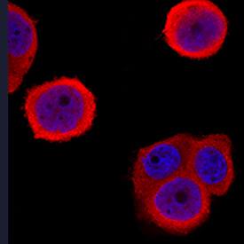
- S100A9 in MDA-MB-468 Human Cell Line. S100A9 was detected in immersion fixed MDA-MB-468 human breast cancer cell line using Sheep Anti-Human S100A9 Antigen Affinity-purified Polyclonal Antibody (Catalog # AF5578) at 1.7 µg/mL for 3 hours at room temperature. Cells were stained using the NorthernLights™ 557-conjugated Anti-Sheep IgG Secondary Antibody (red; Catalog # NL010) and counterstained with DAPI (blue). Specific staining was localized to cytoplasm. View our protocol for Fluorescent ICC Staining of Cells on Coverslips.
Supportive validation
- Submitted by
- R&D Systems (provider)
- Main image

- Experimental details
- S100A9 in Human Cartilage. S100A9 was detected in immersion fixed paraffin-embedded sections of human cartilage using Sheep Anti-Human S100A9 Antigen Affinity-purified Polyclonal Antibody (Catalog # AF5578) at 1 µg/mL overnight at 4 °C. Tissue was stained using the Anti-Sheep HRP-DAB Cell & Tissue Staining Kit (brown; Catalog # CTS019) and counterstained with hematoxylin (blue). Specific staining was localized to cytoplasm of chondrocytes. View our protocol for Chromogenic IHC Staining of Paraffin-embedded Tissue Sections.

Supportive validation
- Submitted by
- R&D Systems (provider)
- Main image

- Experimental details
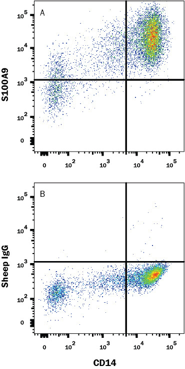
- Detection of S100A9 in Human PBMCs by Flow Cytometry. Human peripheral blood monocytes (PBMC) were stained with Mouse Anti-Human CD14 PE-conjugated Monoclonal Antibody (Catalog # FAB3832P) and either (A) Sheep Anti-Human S100A9 Polyclonal Antibody (Catalog # AF5578) or (B) Sheep IgG Isotype Control (Catalog # 5-001-A) followed by anti-Sheep IgG APC-conjugated Secondary Antibody (Catalog # F0127). To facilitate intracellular staining, cells were fixed with Flow Cytometry Fixation Buffer (Catalog # FC004) and permeabilized with Flow Cytometry Permeabilization/Wash Buffer (Catalog # FC005). View our protocol for Staining Membrane-associated Proteins.