Explore
 Validate
 Learn
Learn Western blot
Antibody data
- Antibody Data
- Antigen structure
- References [0]
- Comments [0]
- Validations
- Western blot [2]
- Immunocytochemistry [2]
- Immunohistochemistry [3]
Submit
Validation data
Reference
Comment
Report error
- Product number
- PA5-118660 - Provider product page

- Provider
- Invitrogen Antibodies
- Product name
- Phospho-PKC theta (Tyr90) Polyclonal Antibody
- Antibody type
- Polyclonal
- Antigen
- Synthetic peptide
- Reactivity
- Human, Mouse, Rat
- Host
- Rabbit
- Isotype
- IgG
- Vial size
- 100 µL
- Concentration
- 1 mg/mL
- Storage
- -20°C
No comments: Submit comment
Supportive validation
- Submitted by
- Invitrogen Antibodies (provider)
- Main image
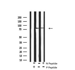
- Experimental details
- Western blot analysis of Phospho-PKC theta (Tyr90) in Jurkat cells using a Phospho-PKC theta (Tyr90) polyclonal antibody (Product # PA5-118660). The -/+ symbols identify the absence or presence of N peptide (non-phospho peptide) and P peptide(phospho peptide).

- Submitted by
- Invitrogen Antibodies (provider)
- Main image
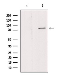
- Experimental details
- Western blot analysis of Phospho-PKC theta (Tyr90) in Hepg2 cells using a Phospho-PKC theta (Tyr90) polyclonal antibody (Product # PA5-118660). Lane 1 was treated with the blocking peptide.

Supportive validation
- Submitted by
- Invitrogen Antibodies (provider)
- Main image
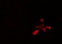
- Experimental details
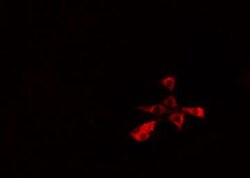
- Immunocytochemical analysis of Phospho-PKC theta (Tyr90) in Hela cells using a Phospho-PKC theta (Tyr90) polyclonal antibody (Product # PA5-118660). The samples were fixed with PFA and permeabilized in 0.1% Triton X-100,then blocked in 10% serum for 45 minutes at 25°C. The primary antibody was diluted at 1:200 and incubated with the sample for 1 hour at 37°C. An Alexa Fluor 594 conjugated goat anti-rabbit IgG (H+L) antibody(Red), diluted at 1:600, was used as secondary antibody.

- Submitted by
- Invitrogen Antibodies (provider)
- Main image
- Experimental details
- Immunocytochemical analysis of Phospho-PKC theta (Tyr90) in Hela cells using a Phospho-PKC theta (Tyr90) polyclonal antibody (Product # PA5-118660). The samples were fixed with PFA and permeabilized in 0.1% Triton X-100,then blocked in 10% serum for 45 minutes at 25°C. The primary antibody was diluted at 1:200 and incubated with the sample for 1 hour at 37°C. An Alexa Fluor 594 conjugated goat anti-rabbit IgG (H+L) antibody(Red), diluted at 1:600, was used as secondary antibody.

Supportive validation
- Submitted by
- Invitrogen Antibodies (provider)
- Main image
- Experimental details
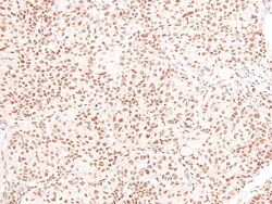
- Immunohistochemical analysis of Phospho-PKC theta (Tyr90) in Human lung cancer tissue using a Phospho-PKC theta (Tyr90) polyclonal antibody (Product # PA5-118660). The tissue was formaldehyde fixed and a heat mediated antigen retrieval step in citrate buffer was performed. The tissue was then blocked and incubated with the antibody for 1.5 hours at 22°C. An HRP conjugated goat anti-rabbit antibody was used as the secondary antibody.

- Submitted by
- Invitrogen Antibodies (provider)
- Main image

- Experimental details
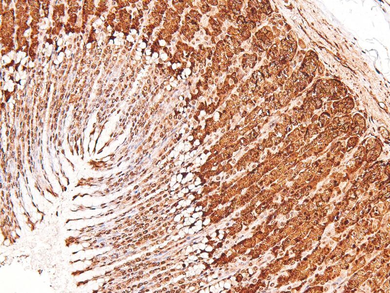
- Immunohistochemical analysis of Phospho-PKC theta (Tyr90) in Rat gastric tissue using a Phospho-PKC theta (Tyr90) polyclonal antibody (Product # PA5-118660). The tissue was formaldehyde fixed and a heat mediated antigen retrieval step in citrate buffer was performed. The tissue was then blocked and incubated with the antibody for 1.5 hours at 22°C. An HRP conjugated goat anti-rabbit antibody was used as the secondary antibody.
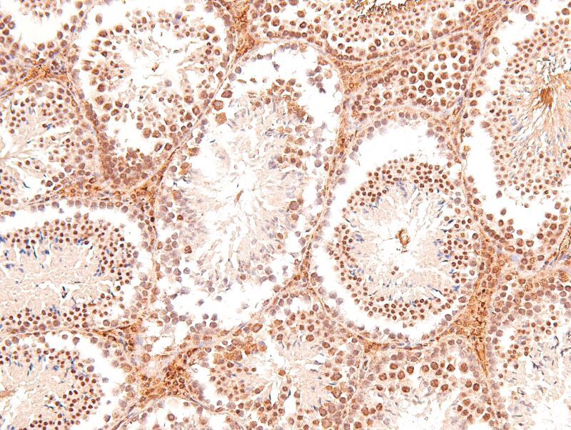
- Submitted by
- Invitrogen Antibodies (provider)
- Main image
- Experimental details
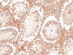
- Immunohistochemical analysis of Phospho-PKC theta (Tyr90) in Mouse testis tissue using a Phospho-PKC theta (Tyr90) polyclonal antibody (Product # PA5-118660). The tissue was formaldehyde fixed and a heat mediated antigen retrieval step in citrate buffer was performed. The tissue was then blocked and incubated with the antibody for 1.5 hours at 22°C. An HRP conjugated goat anti-rabbit antibody was used as the secondary antibody.