 Explore
 Validate
 Learn
Learn Western blot
 Immunocytochemistry
Antibody data
- Antibody Data
- Antigen structure
- References [1]
- Comments [0]
- Validations
- Immunocytochemistry [1]
Submit
Validation data
Reference
Comment
Report error
- Product number
- BAF1740 - Provider product page

- Provider
- R&D Systems
- Product name
- Mouse Integrin alpha 2/CD49b Biotinylated Antibody
- Antibody type
- Polyclonal
- Description
- Antigen Affinity-purified. Detects mouse Integrin alpha 2/CD49b in Western blots. In Western blots, approximately 5% cross-reactivity with recombinant human Integrin alpha M is observed and less than 1% cross-reactivity with recombinant mouse (rm) Integrin alpha 4, rmIntegrin alpha 5, and rmIntegrin alpha E is observed.
- Reactivity
- Mouse
- Host
- Sheep
- Conjugate
- Biotin
- Antigen sequence
Q62469- Isotype
- IgG
- Vial size
- 50 ug
- Concentration
- LYOPH
- Storage
- Use a manual defrost freezer and avoid repeated freeze-thaw cycles. 12 months from date of receipt, -20 to -70 °C as supplied. 1 month, 2 to 8 °C under sterile conditions after reconstitution. 6 months, -20 to -70 °C under sterile conditions after reconstitution.
Submitted references Indoleamine 2,3-dioxygenase promotes peritoneal dissemination of ovarian cancer through inhibition of natural killercell function and angiogenesis promotion.
Nonaka H, Saga Y, Fujiwara H, Akimoto H, Yamada A, Kagawa S, Takei Y, Machida S, Takikawa O, Suzuki M
International journal of oncology 2011 Jan;38(1):113-20
International journal of oncology 2011 Jan;38(1):113-20
No comments: Submit comment
Supportive validation
- Submitted by
- R&D Systems (provider)
- Main image
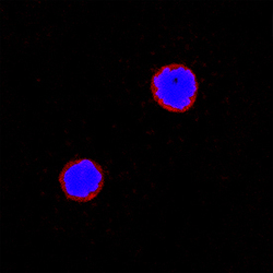
- Experimental details
- Integrin alpha 2/CD49b in Mouse Splenocytes. Integrin alpha 2/CD49b was detected in immersion fixed mouse splenocytes using Sheep Anti-Mouse Integrin alpha 2/CD49b Biotinylated Antigen Affinity-purified Polyclonal Antibody (Catalog # BAF1740) at 15 µg/mL for 3 hours at room temperature. Cells were stained using the NorthernLights™ 557-conjugated Streptavidin (red; Catalog # NL999) and counterstained with DAPI (blue). Specific staining was localized to cell surfaces. View our protocol for Fluorescent ICC Staining of Non-adherent Cells.
