Explore
 Validate
 Learn
Learn Western blot
 Immunohistochemistry
Antibody data
- Antibody Data
- Antigen structure
- References [0]
- Comments [0]
- Validations
- Western blot [1]
- Immunohistochemistry [5]
Submit
Validation data
Reference
Comment
Report error
- Product number
- AMAb91469 - Provider product page

- Provider
- Atlas Antibodies
- Proper citation
- Atlas Antibodies Cat#AMAb91469, RRID:AB_2732114
- Product name
- Anti-ITGA2
- Antibody type
- Monoclonal
- Reactivity
- Human
- Host
- Mouse
- Conjugate
- Unconjugated
- Antigen sequence
ALEAYSETAKVFSIPFHKDCGEDGLCISDLVLDVR
QIPAAQEQPFIVSNQNKRLTFSVTLKNKRESAYNT
GIVVDFSENLFFASFSLPV- Epitope
- Binds to an epitope located within the peptide sequence GEDGLCISDLVLDVR as determined by overlapping synthetic peptides.
- Isotype
- IgG
- Antibody clone number
- CL7318
- Vial size
- 100 µl
- Storage
- Store at +4°C for short term storage. Long time storage is recommended at -20°C.
No comments: Submit comment
Supportive validation
- Submitted by
- Atlas Antibodies (provider)
- Main image

- Experimental details
- Western blot analysis in human cell line RT-4.

Enhanced validation
Supportive validation
- Submitted by
- Atlas Antibodies (provider)
- Enhanced method
- Orthogonal validation
- Main image
- Experimental details
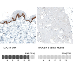
- Immunohistochemistry analysis in human skin and skeletal muscle tissues using AMAb91469 antibody. Corresponding ITGA2 RNA-seq data are presented for the same tissues.
- Sample type
- HUMAN

Supportive validation
- Submitted by
- Atlas Antibodies (provider)
- Main image
- Experimental details
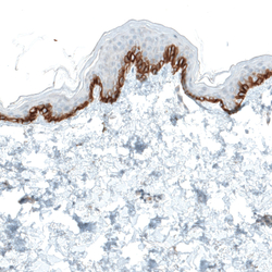
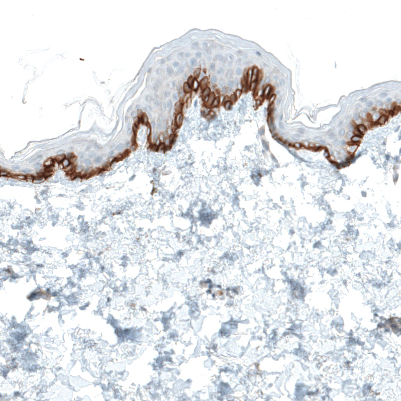
- Immunohistochemical staining of human skin shows strong membranous positivity in cells in basal layer of epidermis.
- Submitted by
- Atlas Antibodies (provider)
- Main image
- Experimental details
- Immunohistochemical staining of human prostate shows moderate membranous positivity in glandular cells.

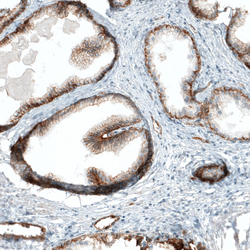
- Submitted by
- Atlas Antibodies (provider)
- Main image
- Experimental details
- Immunohistochemical staining of human small intestine shows moderate to strong membranous positivity in glandular cells.

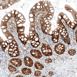
- Submitted by
- Atlas Antibodies (provider)
- Main image
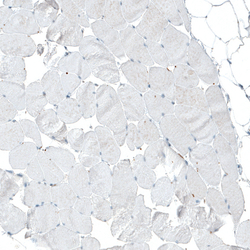
- Experimental details
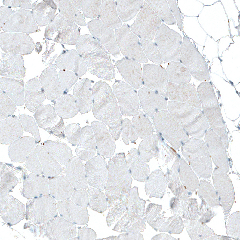
- Immunohistochemical staining of human skeletal muscle shows no positivity in striated muscle fibers as expected.