Explore
 Validate
 Learn
Learn Western blot
 ELISA
ELISAAntibody data
- Antibody Data
- Antigen structure
- References [6]
- Comments [0]
- Validations
- Western blot [1]
- Immunohistochemistry [1]
- Flow cytometry [2]
Submit
Validation data
Reference
Comment
Report error
- Product number
- NBP1-36727 - Provider product page

- Provider
- Novus Biologicals
- Proper citation
- Novus Cat#NBP1-36727, RRID:AB_2197470
- Product name
- Mouse Monoclonal Sulfatase-2/SULF2 Antibody
- Antibody type
- Monoclonal
- Description
- Protein G purified. Does not recognize human or mouse Sulfatase 1 or other family members.
- Reactivity
- Human, Mouse
- Host
- Mouse
- Isotype
- IgG
- Vial size
- 0.1 ml
- Concentration
- 1 mg/ml
- Storage
- Store at 4C short term. Aliquot and store at -20C long term. Avoid freeze-thaw cycles.
Submitted references Epithelial Deletion of Sulf2 Exacerbates Bleomycin-Induced Lung Injury, Inflammation, and Mortality.
Sulfatase 2 promotes breast cancer progression through regulating some tumor-related factors.
MYCN-dependent expression of sulfatase-2 regulates neuroblastoma cell survival.
Overexpression of Sulf2 in idiopathic pulmonary fibrosis.
SULF2 expression by immunohistochemistry and overall survival in oesophageal cancer: a cohort study.
Sulf-2, a heparan sulfate endosulfatase, promotes human lung carcinogenesis.
Yue X
American journal of respiratory cell and molecular biology 2017 Nov;57(5):560-569
American journal of respiratory cell and molecular biology 2017 Nov;57(5):560-569
Sulfatase 2 promotes breast cancer progression through regulating some tumor-related factors.
Zhu C, He L, Zhou X, Nie X, Gu Y
Oncology reports 2016 Mar;35(3):1318-28
Oncology reports 2016 Mar;35(3):1318-28
MYCN-dependent expression of sulfatase-2 regulates neuroblastoma cell survival.
Solari V, Borriello L, Turcatel G, Shimada H, Sposto R, Fernandez GE, Asgharzadeh S, Yates EA, Turnbull JE, DeClerck YA
Cancer research 2014 Nov 1;74(21):5999-6009
Cancer research 2014 Nov 1;74(21):5999-6009
Overexpression of Sulf2 in idiopathic pulmonary fibrosis.
Yue X, Lu J, Auduong L, Sides MD, Lasky JA
Glycobiology 2013 Jun;23(6):709-19
Glycobiology 2013 Jun;23(6):709-19
SULF2 expression by immunohistochemistry and overall survival in oesophageal cancer: a cohort study.
Lui NS, van Zante A, Rosen SD, Jablons DM, Lemjabbar-Alaoui H
BMJ open 2012;2(6)
BMJ open 2012;2(6)
Sulf-2, a heparan sulfate endosulfatase, promotes human lung carcinogenesis.
Lemjabbar-Alaoui H, van Zante A, Singer MS, Xue Q, Wang YQ, Tsay D, He B, Jablons DM, Rosen SD
Oncogene 2010 Feb 4;29(5):635-46
Oncogene 2010 Feb 4;29(5):635-46
No comments: Submit comment
Supportive validation
- Submitted by
- Novus Biologicals (provider)
- Main image

- Experimental details
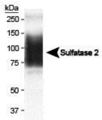
- Western Blot: Sulfatase-2/SULF2 Antibody (2B4) [NBP1-36727] - Analysis showing Sulfatase 2 expression in MCF7 conditioned media (CM). In CM, the 75 kDa band is dominant (Tang R, Rosen SD. J Biol Chem. 2009 Aug 7;284(32):21505-14). Image courtesy of Mark Singer at UCSF.
Supportive validation
- Submitted by
- Novus Biologicals (provider)
- Main image
- Experimental details
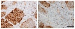
- Immunohistochemistry-Paraffin: Sulfatase-2/SULF2 Antibody (2B4) [NBP1-36727] - Immunohistochemical analysis of Sulfatase 2 in non-small-cell lung carcinoma. (left) lung squamous cell carcinoma stained with 2B4 antibody and (right) tumor-associated stromal cells stained with 2B4 antibody. Image courtesy of Mark Singer at UCSF.

Supportive validation
- Submitted by
- Novus Biologicals (provider)
- Main image
- Experimental details
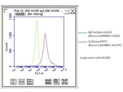
- Flow Cytometry: Sulfatase-2/SULF2 Antibody (2B4) [NBP1-36727] - Analysis using the FITC conjugate of NBP1-36727. Staining of Human lung cancer line with Sulfatase2-FITC. Image from verified customer review.

- Submitted by
- Novus Biologicals (provider)
- Main image
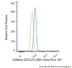
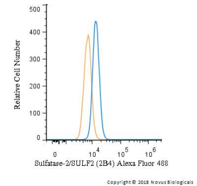
- Experimental details
- Flow Cytometry: Sulfatase-2/SULF2 Antibody (2B4) [NBP1-36727] - An intracellular stain was performed on HeLa cells with Sulfatase-2/SULF2 Antibody (2B4) NBP1-36727AF488 and a matched isotype control (orange). Cells were fixed with 4% PFA and then permeabilized with 0.1% saponin. Cells were incubated in an antibody dilution of 10 ug/mL for 30 minutes at room temperature. Both antibodies were conjugated to Alexa Fluor 488.