PA5-25979
antibody from Invitrogen Antibodies
Targeting: FGFR1
BFGFR, CD331, CEK, FLG, FLT2, H2, H3, H4, H5, KAL2, N-SAM
Antibody data
- Antibody Data
- Antigen structure
- References [1]
- Comments [0]
- Validations
- Western blot [1]
- Immunohistochemistry [1]
- Flow cytometry [1]
- Other assay [1]
Submit
Validation data
Reference
Comment
Report error
- Product number
- PA5-25979 - Provider product page

- Provider
- Invitrogen Antibodies
- Product name
- FGFR1 Polyclonal Antibody
- Antibody type
- Polyclonal
- Antigen
- Other
- Reactivity
- Human, Mouse
- Host
- Rabbit
- Isotype
- IgG
- Vial size
- 400 µL
- Concentration
- 2 mg/mL
- Storage
- Store at 4°C short term. For long term storage, store at -20°C, avoiding freeze/thaw cycles.
Submitted references Calcium and vitamin-D deficiency marginally impairs fracture healing but aggravates posttraumatic bone loss in osteoporotic mice.
Fischer V, Haffner-Luntzer M, Prystaz K, Vom Scheidt A, Busse B, Schinke T, Amling M, Ignatius A
Scientific reports 2017 Aug 3;7(1):7223
Scientific reports 2017 Aug 3;7(1):7223
No comments: Submit comment
Supportive validation
- Submitted by
- Invitrogen Antibodies (provider)
- Main image

- Experimental details
- Western blot analysis using a FGFR1 polyclonal antibody (Product # PA5-25979) in mouse liver tissue lysates (35 µg per lane).
Supportive validation
- Submitted by
- Invitrogen Antibodies (provider)
- Main image

- Experimental details
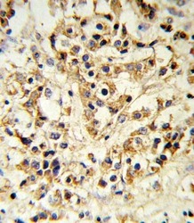
- Immunohistochemistry analysis in formalin-fixed, paraffin-embedded human breast carcinoma using a FGFR1 polyclonal antibody (Product # PA5-25979), followed by HRP-conjugated secondary antibody and DAB staining.
Supportive validation
- Submitted by
- Invitrogen Antibodies (provider)
- Main image

- Experimental details
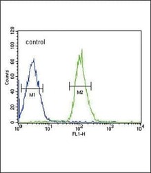
- Flow cytometry analysis of MCF-7 cells using a FGFR1 polyclonal antibody (Product # PA5-25979) (bottom) compared to a negative control cell (top) at a dilution of 1:10-50, followed by a FITC-conjugated goat anti-rabbit antibody
Supportive validation
- Submitted by
- Invitrogen Antibodies (provider)
- Main image

- Experimental details
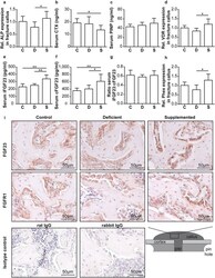
- Figure 4 Serum analysis and gene expression and immunohistochemical analyses of the fracture calli of ovariectomized fractured mice fed a control (C), Ca/VitD-deficient (D) or Ca/VitD-supplemented (S) diet on day 23. ( a ) Relative alkaline phosphatase (ALP) gene expression in the fracture calli (n = 4-5/group). ( b ) Serum C-terminal telopeptide of type I collagen (CTX), and ( c ) serum N-terminal propeptide of type I procollagen (PINP) levels (n = 6-7/group). ( d ) Relative vitamin D-receptor (VDR) gene expression in the fracture calli (n = 4-5/group). ( e ) Serum fibroblast intact growth factor 23 (iFGF23) and ( f ) C-terminal FGF23 (cFGF23) levels (n = 5-7/group). ( g ) Ratio of serum iFGF23 to serum cFGF23 levels (n = 5/group). ( h ) Relative phosphate-regulating neutral endopeptidase, X-linked (Phex) gene expression in the fracture calli (n = 4-5/group). ( i ) Representative images of FGF23, fibroblast growth factor receptor 1 (FGFR1) and species-specific isotype control (rat and rabbit IgG) immunostained sections of the fracture calli. For gene expression analyses, beta-2-microglobulin was used as the house-keeping gene. Data presented as the mean +- SD. Significant differences between C, D and S evaluated by ANOVA/Fishers LSD post-hoc : *p < 0.05, **p < 0.01.
 Explore
 Validate
 Learn
Learn Western blot