34-2000
antibody from Invitrogen Antibodies
Targeting: FZR1
CDC20C, CDH1, FZR, FZR2, HCDH, HCDH1, KIAA1242
Antibody data
- Antibody Data
- Antigen structure
- References [15]
- Comments [0]
- Validations
- Other assay [17]
Submit
Validation data
Reference
Comment
Report error
- Product number
- 34-2000 - Provider product page

- Provider
- Invitrogen Antibodies
- Product name
- FZR1 Polyclonal Antibody
- Antibody type
- Polyclonal
- Antigen
- Synthetic peptide
- Reactivity
- Human, Mouse
- Host
- Rabbit
- Isotype
- IgG
- Vial size
- 100 µg
- Concentration
- 0.25 mg/mL
- Storage
- -20°C
Submitted references Coiled-Coil Domain-Containing 68 Downregulation Promotes Colorectal Cancer Cell Growth by Inhibiting ITCH-Mediated CDK4 Degradation.
CRL4(AMBRA1) is a master regulator of D-type cyclins.
E3 Ubiquitin Ligase APC/C(Cdh1) Regulation of Phenylalanine Hydroxylase Stability and Function.
E3 Ubiquitin Ligase APC/C(Cdh1) Negatively Regulates FAH Protein Stability by Promoting Its Polyubiquitination.
Cdh1 inhibits WWP2-mediated ubiquitination of PTEN to suppress tumorigenesis in an APC-independent manner.
SCF(β-TRCP) promotes cell growth by targeting PR-Set7/Set8 for degradation.
The master cell cycle regulator APC-Cdc20 regulates ciliary length and disassembly of the primary cilium.
The master cell cycle regulator APC-Cdc20 regulates ciliary length and disassembly of the primary cilium.
Regulation of APC(Cdh1) E3 ligase activity by the Fbw7/cyclin E signaling axis contributes to the tumor suppressor function of Fbw7.
SCF-mediated Cdh1 degradation defines a negative feedback system that coordinates cell-cycle progression.
Cdh1 regulates osteoblast function through an APC/C-independent modulation of Smurf1.
Degradation of cyclin A is regulated by acetylation.
Phosphorylation by Akt1 promotes cytoplasmic localization of Skp2 and impairs APCCdh1-mediated Skp2 destruction.
GSK-3 beta targets Cdc25A for ubiquitin-mediated proteolysis, and GSK-3 beta inactivation correlates with Cdc25A overproduction in human cancers.
Cdh1/Hct1-APC is essential for the survival of postmitotic neurons.
Wang C, Li H, Wu L, Jiao X, Jin Z, Zhu Y, Fang Z, Zhang X, Huang H, Zhao L
Frontiers in oncology 2021;11:668743
Frontiers in oncology 2021;11:668743
CRL4(AMBRA1) is a master regulator of D-type cyclins.
Simoneschi D, Rona G, Zhou N, Jeong YT, Jiang S, Milletti G, Arbini AA, O'Sullivan A, Wang AA, Nithikasem S, Keegan S, Siu Y, Cianfanelli V, Maiani E, Nazio F, Cecconi F, Boccalatte F, Fenyö D, Jones DR, Busino L, Pagano M
Nature 2021 Apr;592(7856):789-793
Nature 2021 Apr;592(7856):789-793
E3 Ubiquitin Ligase APC/C(Cdh1) Regulation of Phenylalanine Hydroxylase Stability and Function.
Tyagi A, Sarodaya N, Kaushal K, Chandrasekaran AP, Antao AM, Suresh B, Rhie BH, Kim KS, Ramakrishna S
International journal of molecular sciences 2020 Nov 28;21(23)
International journal of molecular sciences 2020 Nov 28;21(23)
E3 Ubiquitin Ligase APC/C(Cdh1) Negatively Regulates FAH Protein Stability by Promoting Its Polyubiquitination.
Kaushal K, Woo SH, Tyagi A, Kim DH, Suresh B, Kim KS, Ramakrishna S
International journal of molecular sciences 2020 Nov 18;21(22)
International journal of molecular sciences 2020 Nov 18;21(22)
Cdh1 inhibits WWP2-mediated ubiquitination of PTEN to suppress tumorigenesis in an APC-independent manner.
Liu J, Wan L, Liu J, Yuan Z, Zhang J, Guo J, Malumbres M, Liu J, Zou W, Wei W
Cell discovery 2016;2:15044
Cell discovery 2016;2:15044
SCF(β-TRCP) promotes cell growth by targeting PR-Set7/Set8 for degradation.
Wang Z, Dai X, Zhong J, Inuzuka H, Wan L, Li X, Wang L, Ye X, Sun L, Gao D, Zou L, Wei W
Nature communications 2015 Dec 15;6:10185
Nature communications 2015 Dec 15;6:10185
The master cell cycle regulator APC-Cdc20 regulates ciliary length and disassembly of the primary cilium.
Wang W, Wu T, Kirschner MW
eLife 2014 Aug 19;3:e03083
eLife 2014 Aug 19;3:e03083
The master cell cycle regulator APC-Cdc20 regulates ciliary length and disassembly of the primary cilium.
Wang W, Wu T, Kirschner MW
eLife 2014 Aug 19;3:e03083
eLife 2014 Aug 19;3:e03083
Regulation of APC(Cdh1) E3 ligase activity by the Fbw7/cyclin E signaling axis contributes to the tumor suppressor function of Fbw7.
Lau AW, Inuzuka H, Fukushima H, Wan L, Liu P, Gao D, Sun Y, Wei W
Cell research 2013 Jul;23(7):947-61
Cell research 2013 Jul;23(7):947-61
SCF-mediated Cdh1 degradation defines a negative feedback system that coordinates cell-cycle progression.
Fukushima H, Ogura K, Wan L, Lu Y, Li V, Gao D, Liu P, Lau AW, Wu T, Kirschner MW, Inuzuka H, Wei W
Cell reports 2013 Aug 29;4(4):803-16
Cell reports 2013 Aug 29;4(4):803-16
Cdh1 regulates osteoblast function through an APC/C-independent modulation of Smurf1.
Wan L, Zou W, Gao D, Inuzuka H, Fukushima H, Berg AH, Drapp R, Shaik S, Hu D, Lester C, Eguren M, Malumbres M, Glimcher LH, Wei W
Molecular cell 2011 Dec 9;44(5):721-33
Molecular cell 2011 Dec 9;44(5):721-33
Degradation of cyclin A is regulated by acetylation.
Mateo F, Vidal-Laliena M, Canela N, Busino L, Martinez-Balbas MA, Pagano M, Agell N, Bachs O
Oncogene 2009 Jul 23;28(29):2654-66
Oncogene 2009 Jul 23;28(29):2654-66
Phosphorylation by Akt1 promotes cytoplasmic localization of Skp2 and impairs APCCdh1-mediated Skp2 destruction.
Gao D, Inuzuka H, Tseng A, Chin RY, Toker A, Wei W
Nature cell biology 2009 Apr;11(4):397-408
Nature cell biology 2009 Apr;11(4):397-408
GSK-3 beta targets Cdc25A for ubiquitin-mediated proteolysis, and GSK-3 beta inactivation correlates with Cdc25A overproduction in human cancers.
Kang T, Wei Y, Honaker Y, Yamaguchi H, Appella E, Hung MC, Piwnica-Worms H
Cancer cell 2008 Jan;13(1):36-47
Cancer cell 2008 Jan;13(1):36-47
Cdh1/Hct1-APC is essential for the survival of postmitotic neurons.
Almeida A, Bolaños JP, Moreno S
The Journal of neuroscience : the official journal of the Society for Neuroscience 2005 Sep 7;25(36):8115-21
The Journal of neuroscience : the official journal of the Society for Neuroscience 2005 Sep 7;25(36):8115-21
No comments: Submit comment
Supportive validation
- Submitted by
- Invitrogen Antibodies (provider)
- Main image
- Experimental details
- NULL
- Submitted by
- Invitrogen Antibodies (provider)
- Main image

- Experimental details
- NULL
- Submitted by
- Invitrogen Antibodies (provider)
- Main image

- Experimental details
- NULL
- Submitted by
- Invitrogen Antibodies (provider)
- Main image
- Experimental details
- NULL
- Submitted by
- Invitrogen Antibodies (provider)
- Main image

- Experimental details
- NULL
- Submitted by
- Invitrogen Antibodies (provider)
- Main image
- Experimental details
- NULL
- Submitted by
- Invitrogen Antibodies (provider)
- Main image
- Experimental details
- NULL
- Submitted by
- Invitrogen Antibodies (provider)
- Main image

- Experimental details
- NULL
- Submitted by
- Invitrogen Antibodies (provider)
- Main image
- Experimental details
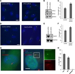
- Figure 2. APC-Cdc20 negatively regulates the length of primary cilia. ( A and D ) RPE1 cells were treated with control siRNA or siRNA targeting APC2 or Cdc20 respectively, serum starved for 48 hr, and stained for acetylated tubulin (green) and DNA (blue). The scale bar represents 10 mum. ( B and E ) Western blot showed knockdown efficiency of siRNA treatment against Cdc20 and APc2. ( C and F ) Ciliary length was measured from ciliated cells based on acetylated-tubulin staining (n > 30). Data are means +-S.D. p
- Submitted by
- Invitrogen Antibodies (provider)
- Main image

- Experimental details
- Figure 5. Nek1 is the substrate of APC-Cdc20 for the regulation of the structure of primary cilia. ( A ) RPE1 cells were treated with control siRNA or Nek1 targeting siRNA, serum starved for 48 hr, and stained for acetylated tubulin (red), gamma-tubulin (green) and DNA (blue). Scale bar represents 5 mum. ( B ) siRNA-treated cells were subjected to Western blot for endogenous Nek1 and GAPDH. ( C ) Cells were transfected with control siRNA, Nek1 siRNA, or Nek1 siRNA together with Cdc20 siRNA respectively, serum starved for 2 days, and stained for acetylated tubulin. The percentage of cells with normal cilia or branched abnormal cilia was calculated from three independent experiments. Data are means +-S.D. ( D ) Western blot showed the decrease of Nek1 protein levels after serum stimulation. ( E ) Degradation of 35 S-labelled Nek1 was examined in cell extracts prepared from HeLa S3 cells. Emi1 was added to test if the degradation was dependent on APC activity. The data were analyzed by autoradiography. ( F ) HA-tagged Nek1 was expressed and labeled with 35 S in reticulocyte lysate. 35 S-labelled Nek1 was purified through HA tag and subjected to in vitro ubiquitylation catalyzed by purified APC. ( G ) RPE1 cells were transfected with plasmid expressing HA-Nek1 and/or Myc-Cdc20, starved for 2 days and subjected to western blot for Myc tag, HA tag, and GAPDH. DOI: http://dx.doi.org/ Figure 5--figure supplement 1. Test of ciliary proteins as APC substrates. ( A ) RPE1 cells were sta
- Submitted by
- Invitrogen Antibodies (provider)
- Main image

- Experimental details
- Figure 1--figure supplement 1. Degradation of securin in RPE1 cell extracts. Cytosolic cell extracts were made from unsynchronized proliferating, serum-starved RPE1 cells respectively. Degradation of securin in these cell extracts at indicated time points was analyzed by western blot. TAME was added to test the dependence of the degradation on APC activity. Cell extract immunodepleted of Cdc20 was used for degradation assay as comparison. DOI: http://dx.doi.org/
- Submitted by
- Invitrogen Antibodies (provider)
- Main image
- Experimental details
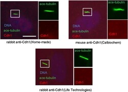
- Figure 1--figure supplement 3. Cdh1 is not localized to the basal body of primary cilia. Starved RPE1 cells were fixed and stained for acetylated tubulin (green), DNA (blue), and Cdh1 (red). For Immunostaining of endogenous Cdh1, three different antibodies against Cdh1 were used as depicted. DOI: http://dx.doi.org/
- Submitted by
- Invitrogen Antibodies (provider)
- Main image
- Experimental details
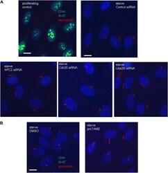
- Figure 2--figure supplement 1. The cells with longer cilia after inhibition of APC-Cdc20 are still in the quiescent stage. ( A ) Proliferating PRE1 cells, starved cells transfected with siRNA against APC2, Cdc20, or Ube2S were fixed and stained for ace-tubulin (red), DNA (blue), and Ki-67 (green, the proliferation marker). ( B ) RPE1 cells were serum-starved for 60 hr and further starved for 3 hr in the presence of DMSO or 20 muM proTAME before staining for acetylated-tubulin (red), Ki-67 (green), and DNA (blue). The scale bars represent 10 mum. DOI: http://dx.doi.org/
- Submitted by
- Invitrogen Antibodies (provider)
- Main image
- Experimental details
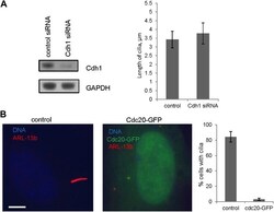
- Figure 2--figure supplement 2. The effects of deregulation of APC co-activators on ciliogenesis. ( A ) RPE1 cells were treated with control siRNA or siRNA-targeting Cdh1 respectively, serum starved for 48 hr. Samples were collected for western blot for Cdh1 and GAPDH. Ciliary length was measured from ciliated cells based on acetylated-tubulin staining (n > 30). Data are means +-S.D. ( B ) RPE1 cells were transiently transfected with control vector or vector expressing Cdc20-GFP (green), serum starved for 48 hr, and stained for Arl13b (red) and DNA (blue). The scale bar represents 5 mum. Percentage of ciliated cells was calculated in control and Cdc20-GFP transfected cells. DOI: http://dx.doi.org/
- Submitted by
- Invitrogen Antibodies (provider)
- Main image

- Experimental details
- Figure 5--figure supplement 1. Test of ciliary proteins as APC substrates. ( A ) RPE1 cells were starved for 48 hr, samples were collected at indicated time point after serum addition, and were subjected to western blot for Aurora-A and GAPDH. ( B ) RPE1 cells were transfected with control siRNA or siRNA against Cdc20, samples were collected after serum starved for 48 hr or 1 hr post serum addition and subjected for western blot for Aurora-A, Cdc20, and GAPDH. ( C ) 35 S-labelled dishevelled protein (DVL) and securin were incubated in the HeLa S3 cell extracts. Samples were collected at indicated times and analyzed by autoradiography. Emi1 was added in the reaction to test whether the degradation was APC dependent. ( D ) 35 S-labelled dishevelled and securin were subjected to in vitro ubiquitylation assay catalyzed by purified APC, recombinant Cdh1 and UBCH10. DOI: http://dx.doi.org/
- Submitted by
- Invitrogen Antibodies (provider)
- Main image

- Experimental details
- Figure 2 Set8 protein stability is negatively controlled by the SCF beta-TRCP1 E3 ligase complex. ( a ) Immunoblot (IB) analysis of whole cell lysates (WCL) derived from HeLa cells infected with the indicated shRNA constructs. ( b ) IB analysis of HeLa cells transfected with the indicated siRNA oligonucleotides. ( c ) IB analysis of WCL derived from HeLa cells infected with shRNA constructs specific for GFP, beta-TRCP1 (six independent lentiviral beta-TRCP1-targeting shRNA constructs namely, -A, -B, -C, -D, -E, -F), or beta-TRCP1+2, followed by selection with 1 mug ml -1 puromycin for 3 days to eliminate non-infected cells. ( d ) IB analysis of WCL from 293 T cells infected with shRNA specific for GFP, or several shRNA constructs against Cullin 1 (three independent lentiviral Cullin 1-targeting shRNA constructs namely, -A, -B, -C) followed by selection with 1 mug ml -1 puromycin for 3 days to eliminate the non-infected cells. ( e ) IB analysis of WCL derived from 293 T cells with or without MG132 treatment. ( f ) HeLa cells were infected with the shGFP or shbeta-TRCP1 followed by selection with 1 mug ml -1 puromycin for 3 days to eliminate non-infected cells. The generated stable cell lines were then treated with nocodazole to arrest at the M phase, and then release back to the cell cycle by washing off nocodazole. At the indicated time points, WCL were prepared and immunoblots were probed with the indicated antibodies. ( g ) HeLa cells were infected with the shRNA constructs
- Submitted by
- Invitrogen Antibodies (provider)
- Main image
- Experimental details
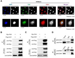
- Figure 5 APC/C Cdh1 co-localizes and interacts with FAH. ( A ) Co-localization of FAH and Cdh1 was performed in HHSteCs by immunostaining with specific antibodies. DAPI was used for nuclear staining (scale bar: 10 um) and the quantification for colocalization coefficients were done using Fiji/ImageJ software. To quantify the degree of co-localization in between the fluorochromes, the Pearson's correlation coefficient (r) was used. The classical Pearson coefficient of the pixel-intensity correlation of colocalization obtained from the coloc2 analysis were mentioned. ( B ) Myc-tagged FAH and Flag-tagged Cdh1 were co-transfected into HEK293 cells. Samples were immunoprecipitated using anti-Myc and immunoblotted using anti-Flag antibodies. GAPDH was used as the loading control. ( C ) Myc-tagged FAH and Flag-tagged Cdh1 were co-transfected into HEK293 cells. Samples were immunoprecipitated using anti-Flag and immunoblotted using anti-Myc antibodies. GAPDH was used as the loading control. ( D ) Endogenous binding between FAH and Cdh1 proteins was performed in HHSteCs. Cell lysates from HHSteCs were immunoprecipitated with specific FAH or Cdh1 antibodies and immunoblotted with FAH or Cdh1 antibodies.
 Explore
 Validate
 Learn
Learn Western blot
 Other assay