Antibody data
- Antibody Data
- Antigen structure
- References [3]
- Comments [0]
- Validations
- Western blot [2]
- Immunocytochemistry [1]
- Immunohistochemistry [3]
- Other assay [2]
Submit
Validation data
Reference
Comment
Report error
- Product number
- PA5-20543 - Provider product page

- Provider
- Invitrogen Antibodies
- Product name
- SLC22A17 Polyclonal Antibody
- Antibody type
- Polyclonal
- Antigen
- Synthetic peptide
- Description
- A suggested positive control is SK-N-SH cell lysate. PA5-20543 can be used with blocking peptide PEP-0663.
- Reactivity
- Human, Mouse, Rat
- Host
- Rabbit
- Isotype
- IgG
- Vial size
- 100 µg
- Concentration
- 1 mg/mL
- Storage
- Maintain refrigerated at 2-8°C for up to 3 months. For long term storage store at -20°C
Submitted references Lipocalin-2 deficiency may predispose to the progression of spontaneous age-related adiposity in mice.
The iron load of lipocalin-2 (LCN-2) defines its pro-tumour function in clear-cell renal cell carcinoma.
Interaction of Alt a 1 with SLC22A17 in the airway mucosa.
Meyers K, López M, Ho J, Wills S, Rayalam S, Taval S
Scientific reports 2020 Sep 3;10(1):14589
Scientific reports 2020 Sep 3;10(1):14589
The iron load of lipocalin-2 (LCN-2) defines its pro-tumour function in clear-cell renal cell carcinoma.
Rehwald C, Schnetz M, Urbschat A, Mertens C, Meier JK, Bauer R, Baer P, Winslow S, Roos FC, Zwicker K, Huard A, Weigert A, Brüne B, Jung M
British journal of cancer 2020 Feb;122(3):421-433
British journal of cancer 2020 Feb;122(3):421-433
Interaction of Alt a 1 with SLC22A17 in the airway mucosa.
Garrido-Arandia M, Tome-Amat J, Pazos-Castro D, Esteban V, Escribese MM, Hernández-Ramírez G, Yuste-Montalvo A, Barber D, Pacios LF, Díaz-Perales A
Allergy 2019 Nov;74(11):2167-2180
Allergy 2019 Nov;74(11):2167-2180
No comments: Submit comment
Supportive validation
- Submitted by
- Invitrogen Antibodies (provider)
- Main image

- Experimental details
- Western blot analysis of Slc22A17 in SK-N-SH lysate with Slc22A17 antibody at 0.5 µg/mL in (A) the presence and (B) the absence of blocking peptide.
- Submitted by
- Invitrogen Antibodies (provider)
- Main image

- Experimental details
- Western Blot analysis of Slc22A17 in SK-N-SH lysate with SLC22A17 Polyclonal Antibody (Product # PA5-20543) at 0.5 µg/mL in (A) the presence and (B) the absence of blocking peptide.
Supportive validation
- Submitted by
- Invitrogen Antibodies (provider)
- Main image

- Experimental details
- Immunofluorescent analysis of rat kidney cells using a Slc22A17 polyclonal antibody (Product # PA5-20543) at a 20 µg/mL dilution.
Supportive validation
- Submitted by
- Invitrogen Antibodies (provider)
- Main image

- Experimental details
- Immunofluorescence of SLC22A17 in mouse kidney tissue with SLC22A17 Polyclonal Antibody (Product # PA5-20543) at 20 µg/mL. Green: Slc22A17 Blue: DAPI staining
- Submitted by
- Invitrogen Antibodies (provider)
- Main image

- Experimental details
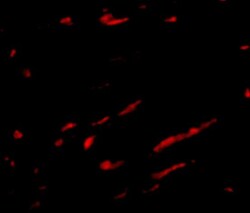
- Immunofluorescence of Slc22A17 in rat kidney tissue tissue with SLC22A17 Polyclonal Antibody (Product # PA5-20543) at 20 µg/mL.
- Submitted by
- Invitrogen Antibodies (provider)
- Main image

- Experimental details
- Immunohistochemistry of SLC22A17 in mouse kidney tissue with SLC22A17 Polyclonal Antibody (Product # PA5-20543) at 2.5 µg/mL.
Supportive validation
- Submitted by
- Invitrogen Antibodies (provider)
- Main image

- Experimental details
- Figure 1 Lcn2 receptor (24p3R) is expressed in 3T3-L1 adipocytes. 3T3-L1 pre-adipocytes were cultured and differentiated to mature adipocytes as described under "" Materials and methods "" section. The Lcn2 receptor (24p3R) expression in 3T3-L1 cells was confirmed by western blot analysis. M markers, UD undifferentiated cells, D differentiated cells. Data shown are representative of two individual experiments. The picture represents the cropped blot and the uncropped image is provided in Supplementary Fig. S1 .
- Submitted by
- Invitrogen Antibodies (provider)
- Main image

- Experimental details
- Fig. 2 LCN-2 protein is elevated in ccRCC. a Healthy and tumour tissue were stained for LCN-2 (green). Nuclear counterstain with DAPI is displayed in white. Representative pictures are given (left), and the quantification of % LCN-2-positive signal was calculated (right). Six pictures of n >=11 patients were quantified, respectively. b Correlation of the LCN-2-positive signal in higher differentiated tumours (G1-G2) and less differentiated tumours (G3-G4). c Percentage of positive cells to lower (pT1-pT2) and higher (pT3-pT4) T stage in comparison with healthy tissue ( n >=11). d LCN-2R mRNA expression normalised to the housekeeping gene 18 S of whole-tissue homogenate of matched renal tumour and adjacent healthy tissue. e Living single cells of healthy and tumour tissue were analysed by FACS. CD45 + immune cells were separated from CD326 + epithelial/tumour cells, which were subsequently analysed for LCN-2R expression, displayed as MFI (mean fluorescence intensity). f Kaplan-Meier curve of high or low LCN-2R expression, the number of patients in brackets. R2 bioinformatics platform was used to probe available TCGA data on ccRCC (KIRC data set). Graphs are displayed as means +- SEM with * p < 0.05.
 Explore
 Validate
 Learn
Learn Western blot