 Explore
 Validate
 Learn
Learn Western blot
Antibody data
- Antibody Data
- Antigen structure
- References [4]
- Comments [0]
- Validations
- Western blot [2]
- Immunocytochemistry [1]
- Other assay [7]
Submit
Validation data
Reference
Comment
Report error
- Product number
- 40-9000 - Provider product page

- Provider
- Invitrogen Antibodies
- Product name
- JAM3 Polyclonal Antibody
- Antibody type
- Polyclonal
- Antigen
- Synthetic peptide
- Reactivity
- Human, Mouse
- Host
- Rabbit
- Isotype
- IgG
- Vial size
- 100 µg
- Concentration
- 0.25 mg/mL
- Storage
- -20°C
Submitted references Soluble JAM-C Ectodomain Serves as the Niche for Adipose-Derived Stromal/Stem Cells.
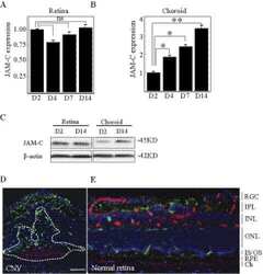
Targeting of junctional adhesion molecule-C inhibits experimental choroidal neovascularization.
Spermatogonial stem cells alone are not sufficient to re-initiate spermatogenesis in the rat testis following adjudin-induced infertility.
Evidence for cross-reactivity of JAM-C antibodies: implications for cellular localization studies.
Yamazaki M, Sugimoto K, Mabuchi Y, Yamashita R, Ichikawa-Tomikawa N, Kaneko T, Akazawa C, Hasegawa H, Imura T, Chiba H
Biomedicines 2021 Mar 10;9(3)
Biomedicines 2021 Mar 10;9(3)
Targeting of junctional adhesion molecule-C inhibits experimental choroidal neovascularization.
Hou X, Hu D, Wang YS, Tang ZS, Zhang F, Chavakis T, Li Y, Li X
Investigative ophthalmology & visual science 2012 Mar;53(3):1584-91
Investigative ophthalmology & visual science 2012 Mar;53(3):1584-91
Spermatogonial stem cells alone are not sufficient to re-initiate spermatogenesis in the rat testis following adjudin-induced infertility.
Mok KW, Mruk DD, Lee WM, Cheng CY
International journal of andrology 2012 Feb;35(1):86-101
International journal of andrology 2012 Feb;35(1):86-101
Evidence for cross-reactivity of JAM-C antibodies: implications for cellular localization studies.
Betanzos A, Schnoor M, Severson EA, Liang TW, Parkos CA
Biology of the cell 2009 Jun 4;101(8):441-53
Biology of the cell 2009 Jun 4;101(8):441-53
No comments: Submit comment
Supportive validation
- Submitted by
- Invitrogen Antibodies (provider)
- Main image
- Experimental details
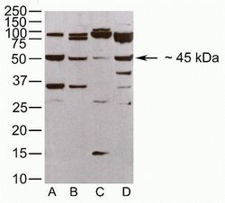
- Western blot analysis of (A) mouse heart and (B) kidney homogenates and (C) EL-4 and (D) NGP96 cell lysates using Zymed Rb anti-JAM-C (Mid) (Product # 40-9000).

- Submitted by
- Invitrogen Antibodies (provider)
- Main image

- Experimental details
- Western blot analysis of (A) mouse heart and (B) kidney homogenates and (C) EL-4 and (D) NGP96 cell lysates using Zymed Rb anti-JAM-C (Mid) (Product # 40-9000).

Supportive validation
- Submitted by
- Invitrogen Antibodies (provider)
- Main image

- Experimental details
- Immunofluorescence analysis of JAM-C was performed using 70% confluent log phase SH-SY5Y cells. The cells were fixed with 4% paraformaldehyde for 10 minutes, permeabilized with 0.1% Triton™ X-100 for 10 minutes, and blocked with 2% BSA for 1 hour at room temperature. The cells were labeled with JAM-C Rabbit Polyclonal Antibody (Product # 40-9000) at 2 µg/mL in 0.1% BSA and incubated for 3 hours at room temperature and then labeled with Goat anti-Rabbit IgG (H+L) Superclonal™ Secondary Antibody, Alexa Fluor® 488 conjugate (Product # A27034 at a dilution of 1:2000 for 45 minutes at room temperature (Panel a: green). Nuclei (Panel b: blue) were stained with SlowFade® Gold Antifade Mountant with DAPI (Product # S36938). F-actin (Panel c: red) was stained with Alexa Fluor® 555 Rhodamine Phalloidin (Product # R415, 1:300). Panel d represents the merged image showing cytoplasmic localization. Panel e shows the no primary antibody control. The images were captured at 60X magnification.

Supportive validation
- Submitted by
- Invitrogen Antibodies (provider)
- Main image
- Experimental details
- NULL

- Submitted by
- Invitrogen Antibodies (provider)
- Main image

- Experimental details
- NULL

- Submitted by
- Invitrogen Antibodies (provider)
- Main image

- Experimental details
- NULL

- Submitted by
- Invitrogen Antibodies (provider)
- Main image

- Experimental details
- NULL

- Submitted by
- Invitrogen Antibodies (provider)
- Main image

- Experimental details
- NULL

- Submitted by
- Invitrogen Antibodies (provider)
- Main image

- Experimental details
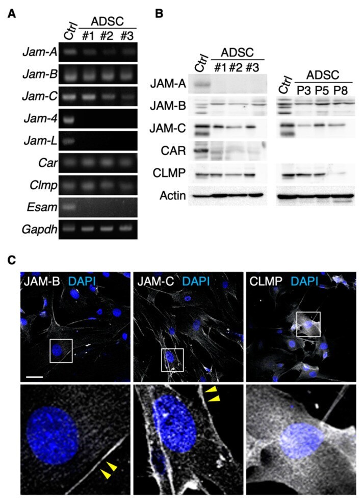
- Figure 1 JAM-B and JAM-C are concentrated on cell membranes of mouse adipose-derived stromal/stem cells (ADSCs). RT-PCR ( A ) and Western blot ( B ) for the indicated molecules in ADSCs derived from three mice (#1-3). Mouse kidney (for Jam-4 ) and spleen tissues (for other junctional adhesion molecules; JAMs) are used as positive controls (Ctrl). P, passage. ( C ) Confocal images of ADSCs stained for the indicated markers. Arrowheads show JAM-B- and JAM-C-immunoreactive signals on the cell membranes of ADSCs. Squares indicate the enlarged areas. Scale bar, 50 um.
- Submitted by
- Invitrogen Antibodies (provider)
- Main image

- Experimental details
- Figure 4 The cleaved JAM-C ectodomain is accumulated in the fat interstitial tissues. ( A ) Schematic illustration for detecting full-length JAM-C (fJAM-C) and/or soluble JAM-C (sJAM-C) using JAM-C (N) and JAM-C (C) antibodies (Abs) targeting the N- and C-termini, respectively. ( B ) Knockdown of the Jam3 gene encoding mouse JAM-C in ADSCs using the CRISPR method. ( C ) Western blot for the indicated proteins in the whole-cell lysates and supernatants of the revealed ADSCs. ( D ) Western blot for the indicated proteins in the whole-cell lysates of mouse adult spleen tissue (control), adipose tissue and ADSCs. ( E ) Confocal images of mouse adipose tissue stained with JAM-C (N) and JAM-C (C) Abs. Asterisks indicate lipid droplets in differentiated adipocytes. Squares show the enlarged areas. Scale bar, 50 um.
