Explore
 Validate
 Learn
Learn Western blot
 Immunohistochemistry
Antibody data
- Antibody Data
- Antigen structure
- References [1]
- Comments [0]
- Validations
- Western blot [4]
- Immunocytochemistry [1]
- Immunohistochemistry [10]
Submit
Validation data
Reference
Comment
Report error
- Product number
- HPA002632 - Provider product page

- Provider
- Atlas Antibodies
- Proper citation
- Atlas Antibodies Cat#HPA002632, RRID:AB_1857316
- Product name
- Anti-SND1
- Antibody type
- Polyclonal
- Reactivity
- Human
- Host
- Rabbit
- Conjugate
- Unconjugated
- Antigen sequence
TLSPAFSTRVLPAQATEYAFAFIQVPQDDDARTDA
VDSVVRDIQNTQCLLNVEHLSAGCPHVTLQFADSK
GDVGLGLVKEGLVMVEVRKEKQFQKVITEYLNAQE
SAKSARLNLWRYGDFRADDADEF- Isotype
- IgG
- Vial size
- 100 µl
- Storage
- Store at +4°C for short term storage. Long time storage is recommended at -20°C.
Submitted references Systematic validation of antibody binding and protein subcellular localization using siRNA and confocal microscopy
Stadler C, Hjelmare M, Neumann B, Jonasson K, Pepperkok R, Uhlén M, Lundberg E
Journal of Proteomics 2012 April;75(7):2236-2251
Journal of Proteomics 2012 April;75(7):2236-2251
No comments: Submit comment
Supportive validation
- Submitted by
- Atlas Antibodies (provider)
- Main image
- Experimental details
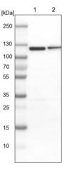
- Lane 1: NIH-3T3 cell lysate (Mouse embryonic fibroblast cells)Lane 2: NBT-II cell lysate (Rat Wistar bladder tumour cells)
- Sample type
- MOUSE, RAT

- Submitted by
- Atlas Antibodies (provider)
- Main image
- Experimental details
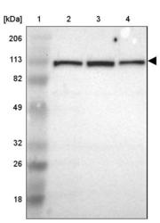
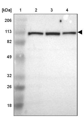
- Lane 1: Marker [kDa] 206, 113, 82, 49, 32, 26, 18Lane 2: Human cell line RT-4Lane 3: Human cell line U-251MG spLane 4: Human cell line A-431
- Sample type
- HUMAN
- Submitted by
- Atlas Antibodies (provider)
- Main image
- Experimental details
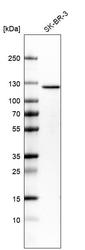
- Western blot analysis in human cell line SK-BR-3.

- Submitted by
- Atlas Antibodies (provider)
- Main image
- Experimental details
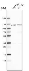
- Western blot analysis in human cell line U-87 MG and human cell line EFO-21.

Supportive validation
- Submitted by
- Atlas Antibodies (provider)
- Main image

- Experimental details
- Immunofluorescent staining of human cell line A-431 shows localization to cytosol.
- Sample type
- HUMAN

Enhanced validation
Enhanced validation
Supportive validation
- Submitted by
- Atlas Antibodies (provider)
- Enhanced method
- Orthogonal validation
- Main image
- Experimental details
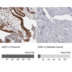
- Immunohistochemistry analysis in human placenta and skeletal muscle tissues using HPA002632 antibody. Corresponding SND1 RNA-seq data are presented for the same tissues.
- Sample type
- HUMAN

Enhanced validation
- Submitted by
- Atlas Antibodies (provider)
- Enhanced method
- Independent antibody validation
- Main image
- Experimental details
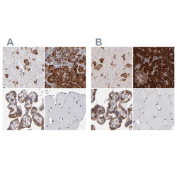
- Immunohistochemical staining of human cerebral cortex, pancreas, placenta and skeletal muscle using Anti-SND1 antibody HPA002632 (A) shows similar protein distribution across tissues to independent antibody HPA002529 (B).

Supportive validation
- Submitted by
- Atlas Antibodies (provider)
- Main image

- Experimental details
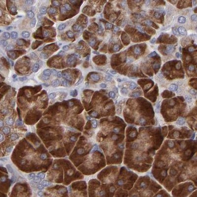
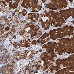
- Immunohistochemical staining of human pancreas shows strong cytoplasmic positivity in exocrine glandular cells.

- Submitted by
- Atlas Antibodies (provider)
- Main image
- Experimental details
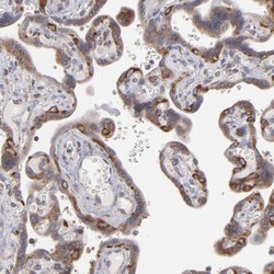
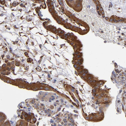
- Immunohistochemical staining of human placenta shows high expression.
- Sample type
- HUMAN

- Submitted by
- Atlas Antibodies (provider)
- Main image
- Experimental details
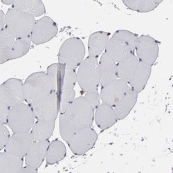
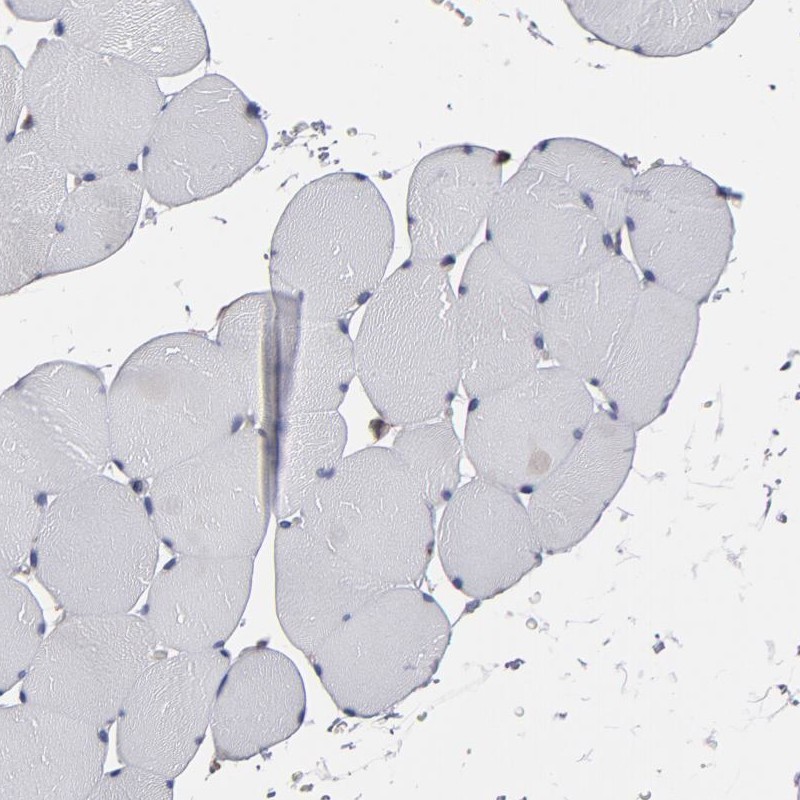
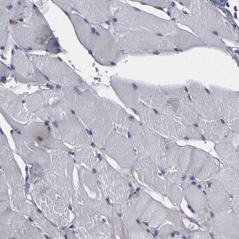
- Immunohistochemical staining of human skeletal muscle shows low expression as expected.
- Sample type
- HUMAN
- Submitted by
- Atlas Antibodies (provider)
- Main image
- Experimental details
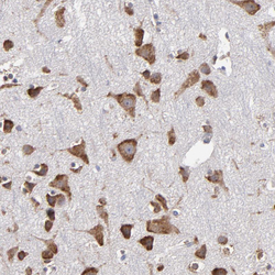
- Immunohistochemical staining of human cerebral cortex shows strong cytoplasmic positivity in neurons.
- Sample type
- HUMAN

- Submitted by
- Atlas Antibodies (provider)
- Main image
- Experimental details
- Immunohistochemical staining of human stomach shows strong cytoplasmic positivity in glandular cells.
- Sample type
- HUMAN

- Submitted by
- Atlas Antibodies (provider)
- Main image
- Experimental details
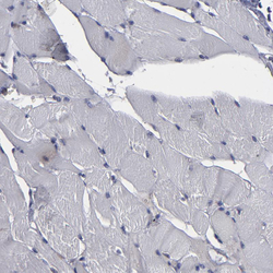
- Immunohistochemical staining of human skeletal muscle shows no positivity in myocytes as expected.
- Sample type
- HUMAN
- Submitted by
- Atlas Antibodies (provider)
- Main image
- Experimental details
- Immunohistochemical staining of human placenta shows strong cytoplasmic positivity in trophoblastic cells.
- Sample type
- HUMAN

- Submitted by
- Atlas Antibodies (provider)
- Main image

- Experimental details
- Immunohistochemical staining of human pancreas using Anti-SND1 antibody HPA002632.
- Sample type
- HUMAN