 Explore
 Validate
 Learn
Learn44-1210G
antibody from Invitrogen Antibodies
Targeting: AURKA
AIK, ARK1, AurA, BTAK, PPP1R47, STK15, STK6, STK7
 Western blot
Antibody data
- Antibody Data
- Antigen structure
- References [2]
- Comments [0]
- Validations
- Western blot [1]
- Immunocytochemistry [1]
- Immunohistochemistry [1]
- Other assay [1]
Submit
Validation data
Reference
Comment
Report error
- Product number
- 44-1210G - Provider product page

- Provider
- Invitrogen Antibodies
- Product name
- Phospho-Aurora A (Thr288) Polyclonal Antibody
- Antibody type
- Polyclonal
- Antigen
- Synthetic peptide
- Reactivity
- Human, Mouse
- Host
- Rabbit
- Isotype
- IgG
- Vial size
- 100 µL
- Storage
- -20°C
Submitted references Novel Aurora A Kinase Inhibitor Fangchinoline Enhances Cisplatin-DNA Adducts and Cisplatin Therapeutic Efficacy in OVCAR-3 Ovarian Cancer Cells-Derived Xenograft Model.
The PP1 regulator PPP1R2 coordinately regulates AURKA and PP1 to control centrosome phosphorylation and maintain central spindle architecture.
Winardi D, Chu PY, Chen GY, Wang K, Hsu WY, Hsieh CL, Chen YH, Wu YC, Yang JC
International journal of molecular sciences 2022 Feb 7;23(3)
International journal of molecular sciences 2022 Feb 7;23(3)
The PP1 regulator PPP1R2 coordinately regulates AURKA and PP1 to control centrosome phosphorylation and maintain central spindle architecture.
Bresch AM, Yerich N, Wang R, Sperry AO
BMC molecular and cell biology 2020 Nov 25;21(1):84
BMC molecular and cell biology 2020 Nov 25;21(1):84
No comments: Submit comment
Supportive validation
- Submitted by
- Invitrogen Antibodies (provider)
- Main image

- Experimental details
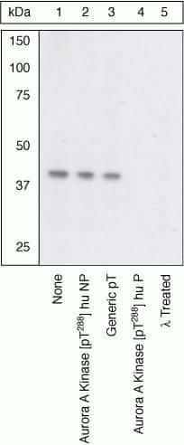
- Peptide Competition. Extracts of serum-starved NIH3T3 cells were resolved by SDS-PAGE on a 10% Tris-glycine gel and transferred to PVDF. The membrane was blocked with a 3% BSA-TBST buffer for one hour at room temperature, and either left untreated (1-4) or treated with lambda phosphatase (5), then incubated with the Aurora A Kinase (pT288) antibody for two hours at room temperature in 3% BSA-TBST buffer, following prior incubation with: no peptide (1, 5), the non-phosphopeptide corresponding to the phosphopeptide immunogen (2), a generic phosphothreonine-containing peptide (3), or the phosphopeptide immunogen (4). After washing, the membrane was incubated with goat F (ab’)2 anti-rabbit IgG HRP conjugate (Product # ALI4404) and signals were detected using the Pierce SuperSignal™ method. The data show that only the phosphopeptide corresponding to Aurora A Kinase (pT288) blocks the antibody signal, and that phosphatase treatment eliminates the signal, thereby demonstrating the specificity of the antibody.
Supportive validation
- Submitted by
- Invitrogen Antibodies (provider)
- Main image

- Experimental details
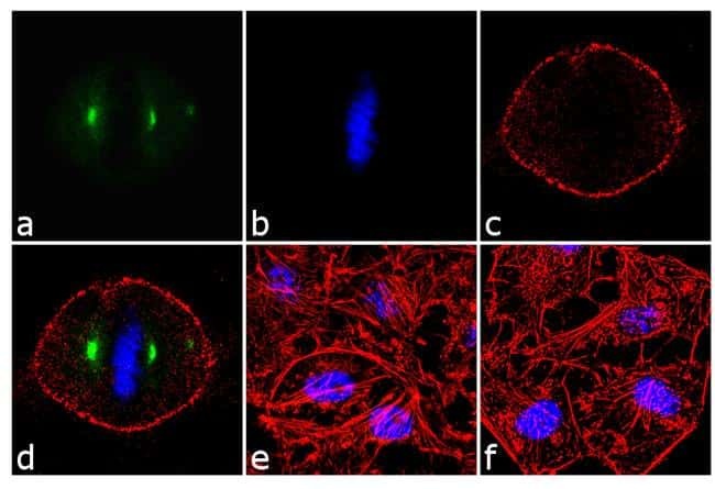
- Immunofluorescence analysis of Phospho-Aurora A pThr288 was done on 70% confluent log phase HeLa cells treated with 3uM Nocodazole for 24hrs. The cells were fixed with 4% paraformaldehyde for 10 minutes, permeabilized with 0.1% Triton™ X-100 for 10 minutes, and blocked with 1% BSA for 1 hour at room temperature. The cells were labeled with Phospho-Aurora A pThr288 Rabbit Polyclonal Antibody (Product # 44-1210G) at 1:250 dilution in 0.1% BSA and incubated for 3 hours at room temperature and then labeled with Goat anti-Rabbit IgG (H+L) Superclonal™ Secondary Antibody, Alexa Fluor® 488 conjugate (Product # A27034) at a dilution of 1:2000 for 45 minutes at room temperature (Panel a: green). Nuclei (Panel b: blue) were stained with SlowFade® Gold Antifade Mountant with DAPI (Product # S36938). F-actin (Panel c: red) was stained with Rhodamine Phalloidin (Product # R415, 1:300). Panel d is a merged image showing spindle localization. Panel e is untreated cell with no signal. Panel f is a no primary antibody control. The images were captured at 60X magnification.
Supportive validation
- Submitted by
- Invitrogen Antibodies (provider)
- Main image

- Experimental details
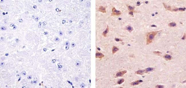
- Immunohistochemistry analysis of Phospho-Aurora A (pThr288) showing staining in the cytoplasm of paraffin-embedded mouse brain tissue (right) compared to a negative control without primary antibody (left). To expose target proteins, antigen retrieval was performed using 10mM sodium citrate (pH 6.0), microwaved for 8-15 min. Following antigen retrieval, tissues were blocked in 3% H2O2-methanol for 15 min at room temperature, washed with ddH2O and PBS, and then probed with a Anti- Phospho-Aurora A (pThr288) Polyclonal Antibody (Product # 44-1210G) diluted in 3% BSA-PBS at a dilution of 1:100 overnight at 4°C in a humidified chamber. Tissues were washed extensively in PBST and detection was performed using an HRP-conjugated secondary antibody followed by colorimetric detection using a DAB kit. Tissues were counterstained with hematoxylin and dehydrated with ethanol and xylene to prep for mounting.
Supportive validation
- Submitted by
- Invitrogen Antibodies (provider)
- Main image
- Experimental details
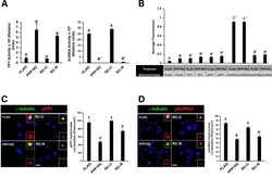
- Fig. 8 The effect of PPP1R2 on PP1 and AURKA activity at the centrosome was dependent on its C-terminus. a A graphical representation of the PP1 and AURKA activity of cells expressing each construct is shown. b Protein expression and phosphorylation levels of proteins indicated by the table below the graph were measured using ELISA. Cells were transfected with the indicated constructs, fixed, and labeled for gamma-tubulin (red) and either ( c ) phosphorylated PP1 (pPP1, green) or ( d ) phosphorylated AURKA (pAURKA, green) antibodies. pPP1 and pAURKA levels at the centrosome were quantified using Metamorph software and are displayed as graphs in C and D. Statistically significant differences ( p
