 Explore
 Validate
 Learn
Learn Western blot
Antibody data
- Antibody Data
- Antigen structure
- References [3]
- Comments [0]
- Validations
- Western blot [3]
- Immunocytochemistry [1]
- Immunohistochemistry [2]
Submit
Validation data
Reference
Comment
Report error
- Product number
- GTX107763 - Provider product page

- Provider
- GeneTex
- Proper citation
- GeneTex Cat#GTX107763, RRID:AB_1951614
- Product name
- Raf1 antibody [N3C3]
- Antibody type
- Polyclonal
- Reactivity
- Human, Mouse
- Host
- Rabbit
Submitted references Melatonin Inhibits the Progression of Hepatocellular Carcinoma through MicroRNA Let7i-3p Mediated RAF1 Reduction.
RAF1 is increased in labouring myometrium and modulates inflammation-induced pro-labour mediators.
SH3BGRL3 Protein as a Potential Prognostic Biomarker for Urothelial Carcinoma: A Novel Binding Partner of Epidermal Growth Factor Receptor.
Wang TH, Hsueh C, Chen CC, Li WS, Yeh CT, Lian JH, Chang JL, Chen CY
International journal of molecular sciences 2018 Sep 10;19(9)
International journal of molecular sciences 2018 Sep 10;19(9)
RAF1 is increased in labouring myometrium and modulates inflammation-induced pro-labour mediators.
Lappas M
Reproduction (Cambridge, England) 2016 Apr;151(4):411-20
Reproduction (Cambridge, England) 2016 Apr;151(4):411-20
SH3BGRL3 Protein as a Potential Prognostic Biomarker for Urothelial Carcinoma: A Novel Binding Partner of Epidermal Growth Factor Receptor.
Chiang CY, Pan CC, Chang HY, Lai MD, Tzai TS, Tsai YS, Ling P, Liu HS, Lee BF, Cheng HL, Ho CL, Chen SH, Chow NH
Clinical cancer research : an official journal of the American Association for Cancer Research 2015 Dec 15;21(24):5601-11
Clinical cancer research : an official journal of the American Association for Cancer Research 2015 Dec 15;21(24):5601-11
No comments: Submit comment
Supportive validation
- Submitted by
- GeneTex (provider)
- Main image
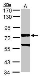
- Experimental details
- Sample (30 ?g of whole cell lysate)A: A431 (GTX27909)7.5% SDS PAGEGTX107763 diluted at 1:5000The HRP-conjugated anti-rabbit IgG antibody (GTX213110-01) was used to detect the primary antibody.
- Submitted by
- GeneTex (provider)
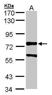
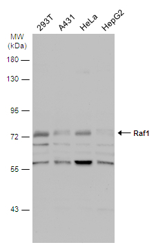
- Main image

- Experimental details
- Various whole cell extracts (30 ?g) were separated by 7.5% SDS-PAGE, and the membrane was blotted with Raf1 antibody [N3C3] (GTX107763) diluted at 1:1000. The HRP-conjugated anti-rabbit IgG antibody (GTX213110-01) was used to detect the primary antibody.
- Submitted by
- GeneTex (provider)
- Main image
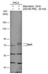
- Experimental details
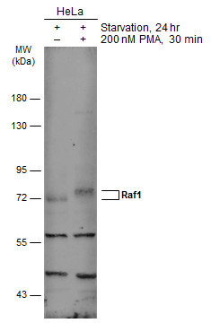
- Untreated (¡V) and treated (+) HeLa whole cell extracts (30 ?g) were separated by 7.5% SDS-PAGE, and the membrane was blotted with Raf1 antibody [N3C3] (GTX107763) diluted at 1:1000. The HRP-conjugated anti-rabbit IgG antibody (GTX213110-01) was used to detect the primary antibody.
Supportive validation
- Submitted by
- GeneTex (provider)
- Main image

- Experimental details
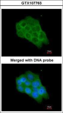
- Immunofluorescence analysis of paraformaldehyde-fixed A431, using Raf1(GTX107763) antibody at 1:200 dilution.
Supportive validation
- Submitted by
- GeneTex (provider)
- Main image
- Experimental details
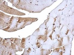
- Raf1 antibody [N3C3] detects Raf1 protein at nucleus on mouse muscle by immunohistochemical analysis. Sample: Paraffin-embedded mouse muscle. Raf1 antibody [N3C3] (GTX107763) dilution: 1:500.

- Submitted by
- GeneTex (provider)
- Main image
- Experimental details
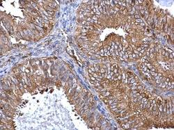
- Raf1 antibody [N3C3] detects Raf1 protein at cytoplasm on human endometrial carcinoma by immunohistochemical analysis. Sample: Paraffin-embedded human endometrial carcinoma. Raf1 antibody [N3C3] (GTX107763) diluted at 1:500.
