GTX109519
antibody from GeneTex
Targeting: MAD1L1
HsMAD1, MAD1, PIG9, TP53I9, TXBP181
 Western blot
 Immunocytochemistry
 Immunoprecipitation
 Immunohistochemistry
 Chromatin Immunoprecipitation
Antibody data
- Antibody Data
- Antigen structure
- References [9]
- Comments [0]
- Validations
- Western blot [5]
- Immunocytochemistry [1]
- Immunoprecipitation [1]
- Immunohistochemistry [1]
Submit
Validation data
Reference
Comment
Report error
- Product number
- GTX109519 - Provider product page

- Provider
- GeneTex
- Proper citation
- GeneTex Cat#GTX109519, RRID:AB_1950847
- Product name
- MAD1 antibody
- Antibody type
- Polyclonal
- Reactivity
- Human, Mouse, Rat
- Host
- Rabbit
Submitted references LMO7 exerts an effect on mitosis progression and the spindle assembly checkpoint.
Spindle assembly checkpoint signaling and sister chromatid cohesion are disrupted by HPV E6-mediated transformation.
Cep57 is a Mis12-interacting kinetochore protein involved in kinetochore targeting of Mad1-Mad2.
Mad1 promotes chromosome congression by anchoring a kinesin motor to the kinetochore.
The RZZ complex requires the N-terminus of KNL1 to mediate optimal Mad1 kinetochore localization in human cells.
Stable kinetochore-microtubule attachment is sufficient to silence the spindle assembly checkpoint in human cells.
Recruitment of the human Cdt1 replication licensing protein by the loop domain of Hec1 is required for stable kinetochore-microtubule attachment.
The spindle checkpoint protein MAD1 regulates the expression of E-cadherin and prevents cell migration.
RED, a spindle pole-associated protein, is required for kinetochore localization of MAD1, mitotic progression, and activation of the spindle assembly checkpoint.
Tzeng YW, Li DY, Chen Y, Yang CH, Chang CY, Juang YL
The international journal of biochemistry & cell biology 2018 Jan;94:22-30
The international journal of biochemistry & cell biology 2018 Jan;94:22-30
Spindle assembly checkpoint signaling and sister chromatid cohesion are disrupted by HPV E6-mediated transformation.
Shirnekhi HK, Kelley EP, DeLuca JG, Herman JA
Molecular biology of the cell 2017 Jul 15;28(15):2035-2041
Molecular biology of the cell 2017 Jul 15;28(15):2035-2041
Cep57 is a Mis12-interacting kinetochore protein involved in kinetochore targeting of Mad1-Mad2.
Zhou H, Wang T, Zheng T, Teng J, Chen J
Nature communications 2016 Jan 8;7:10151
Nature communications 2016 Jan 8;7:10151
Mad1 promotes chromosome congression by anchoring a kinesin motor to the kinetochore.
Akera T, Goto Y, Sato M, Yamamoto M, Watanabe Y
Nature cell biology 2015 Sep;17(9):1124-33
Nature cell biology 2015 Sep;17(9):1124-33
The RZZ complex requires the N-terminus of KNL1 to mediate optimal Mad1 kinetochore localization in human cells.
Caldas GV, Lynch TR, Anderson R, Afreen S, Varma D, DeLuca JG
Open biology 2015 Nov;5(11)
Open biology 2015 Nov;5(11)
Stable kinetochore-microtubule attachment is sufficient to silence the spindle assembly checkpoint in human cells.
Tauchman EC, Boehm FJ, DeLuca JG
Nature communications 2015 Dec 1;6:10036
Nature communications 2015 Dec 1;6:10036
Recruitment of the human Cdt1 replication licensing protein by the loop domain of Hec1 is required for stable kinetochore-microtubule attachment.
Varma D, Chandrasekaran S, Sundin LJ, Reidy KT, Wan X, Chasse DA, Nevis KR, DeLuca JG, Salmon ED, Cook JG
Nature cell biology 2012 May 13;14(6):593-603
Nature cell biology 2012 May 13;14(6):593-603
The spindle checkpoint protein MAD1 regulates the expression of E-cadherin and prevents cell migration.
Chen Y, Yeh PC, Huang JC, Yeh CC, Juang YL
Oncology reports 2012 Feb;27(2):487-91
Oncology reports 2012 Feb;27(2):487-91
RED, a spindle pole-associated protein, is required for kinetochore localization of MAD1, mitotic progression, and activation of the spindle assembly checkpoint.
Yeh PC, Yeh CC, Chen YC, Juang YL
The Journal of biological chemistry 2012 Apr 6;287(15):11704-16
The Journal of biological chemistry 2012 Apr 6;287(15):11704-16
No comments: Submit comment
Supportive validation
- Submitted by
- GeneTex (provider)
- Main image

- Experimental details
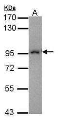
- Sample (30 ?g of whole cell lysate)A: Molt-4 (GTX27912)7.5% SDS PAGEGTX109519 diluted at 1:10000The HRP-conjugated anti-rabbit IgG antibody (GTX213110-01) was used to detect the primary antibody.
- Submitted by
- GeneTex (provider)
- Main image

- Experimental details
- MAD1 antibody detects MAD1L1 protein by western blot analysis.A. 30 ?g Neuro2A whole cell lysate/extract B. 30 ?g GL261 whole cell lysate/extract C. 30 ?g C8D30 whole cell lysate/extract D. 30 ?g NIH-3T3 whole cell lysate/extract E. 30 ?g BCL-1 whole cell lysate/extract F. 30 ?g Raw264.7 whole cell lysate/extract 7.5% SDS-PAGEMAD1 antibody (GTX109519) dilution: 1:3000 The HRP-conjugated anti-rabbit IgG antibody (GTX213110-01) was used to detect the primary antibody.
- Submitted by
- GeneTex (provider)
- Main image

- Experimental details
- MAD1 antibody detects MAD1L1 protein by western blot analysis.A. 30 ?g PC-12 whole cell lysate/extract B. 30 ?g Rat2 whole cell lysate/extract7.5% SDS-PAGEMAD1 antibody (GTX109519) dilution: 1:3000 The HRP-conjugated anti-rabbit IgG antibody (GTX213110-01) was used to detect the primary antibody.
- Submitted by
- GeneTex (provider)
- Main image

- Experimental details
- MAD1 antibody detects MAD1 protein by western blot analysis. Various whole cell extracts (30 ?g) were separated by 7.5% SDS-PAGE, and the membrane was blotted with MAD1 antibody (GTX109519) diluted by 1:5000. The HRP-conjugated anti-rabbit IgG antibody (GTX213110-01) was used to detect the primary antibody.
- Submitted by
- GeneTex (provider)
- Main image

- Experimental details
- MAD1 antibody detects MAD1 protein by Western blot analysis. Various whole cell extracts (30 £gg) were separated by 7.5% SDS-PAGE, and the membrane was blotted with MAD1 antibody (GTX109519) diluted by 1:5000.
Supportive validation
- Submitted by
- GeneTex (provider)
- Main image

- Experimental details
- MAD1 antibody detects MAD1 protein at cytoplasm and nucleus by immunofluorescent analysis.Sample: HeLa cells were fixed in 4% paraformaldehyde at RT for 15 min.Green: MAD1 protein stained by MAD1 antibody (GTX109519) diluted at 1:500.Red: HEC1, a kinetochore marker, stained by HEC1 antibody [9G3.23] (GTX70268) diluted at 1:500.Blue: Hoechst 33342 staining.
Supportive validation
- Submitted by
- GeneTex (provider)
- Main image

- Experimental details
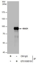
- Immunoprecipitation of MAD1 protein from HeLa whole cell extracts using 5 £gg of MAD1 antibody (GTX109519).Western blot analysis was performed using MAD1 antibody (GTX109519).EasyBlot anti-Rabbit IgG (GTX221666-01) was used as a secondary reagent.
Supportive validation
- Submitted by
- GeneTex (provider)
- Main image

- Experimental details
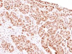
- Immunohistochemical analysis of paraffin-embedded SW480 Xenograft, using Mad 1 (GTX109519) antibody at 1:100 dilution.
 Explore
 Validate
 Learn
Learn