Explore
 Validate
 Learn
Learn Western blot
 Immunohistochemistry
Antibody data
- Antibody Data
- Antigen structure
- References [0]
- Comments [0]
- Validations
- Immunohistochemistry [1]
- Flow cytometry [1]
Submit
Validation data
Reference
Comment
Report error
- Product number
- MAB361-100 - Provider product page

- Provider
- Novus Biologicals
- Product name
- Mouse Monoclonal CCL19/MIP-3 beta Antibody
- Antibody type
- Monoclonal
- Description
- Protein A or G purified from ascites. Detects human CCL19/MIP-3 beta in direct ELISAs and Western blots. In direct ELISAs, no cross-reactivity with recombinant human CCL1, 2, 3, 4, 5, 7, 8, 11, 13, 14, 15, 16, 17, 18, 20, 21, 22, 23, 24, 25, recombinant mouse CCL2, 3, 4, 5, 6, 7, 9, 11, 12, 21, 22, or 25 is observed.
- Reactivity
- Human
- Host
- Mouse
- Conjugate
- Unconjugated
- Isotype
- IgG
- Vial size
- 100 ug
- Storage
- Use a manual defrost freezer and avoid repeated freeze-thaw cycles. 12 months from date of receipt, -20 to -70 degreesC as supplied. 1 month, 2 to 8 degreesC under sterile conditions after reconstitution. 6 months, -20 to -70 degreesC under sterile conditions after reconstitution.
No comments: Submit comment
Supportive validation
- Submitted by
- Novus Biologicals (provider)
- Main image
- Experimental details
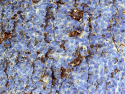
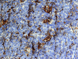
- CCL19/MIP-3 beta in Human Tonsil. CCL19/MIP-3 beta was detected in immersion fixed paraffin-embedded sections of human tonsil using 25 µg/mL Mouse Anti-Human CCL19/ MIP-3 beta Monoclonal Antibody (Catalog # MAB361) overnight at 4 °C. Tissue was stained with the Anti-Mouse HRP-DAB Cell & Tissue Staining Kit (brown; Catalog # CTS002) and counter-stained with hematoxylin (blue). View our protocol for Chromogenic IHC Staining of Paraffin-embedded Tissue Sections.
Supportive validation
- Submitted by
- Novus Biologicals (provider)
- Main image
- Experimental details
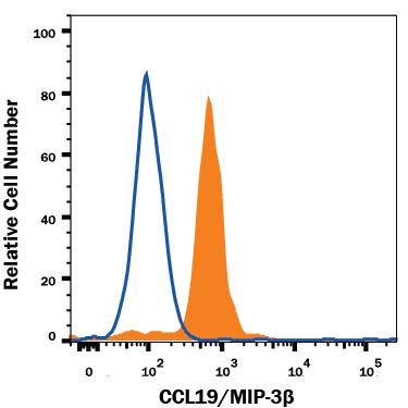
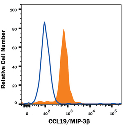
- Detection of CCL19/MIP-3 beta in Human Dendritic Cells by Flow Cytometry. Human monocyte-derived dendritic cells were stained with Mouse Anti-Human CCL19/MIP-3 beta Monoclonal Antibody (Catalog # MAB361, filled histogram) or isotype control antibody (Catalog # MAB0041, open histogram), followed by Phycoerythrin-conjugated Anti-Mouse IgG Secondary Antibody (Catalog # F0102B). To facilitate intracellular staining, cells were fixed with Flow Cytometry Fixation Buffer (Catalog # FC004) and permeabilized with Flow Cytometry Permeabilization/Wash Buffer I (Catalog # FC005).